FPR1 channel function-based chemical carcinogenicity in-vitro detection method
An in vitro detection and chemical technology, applied in biochemical equipment and methods, microbial determination/inspection, material analysis by optical means, etc., can solve the problems of complex and cumbersome direct detection methods and high requirements for experimental environment, and achieve results. Objectively quantifiable, reducing interference factors and shortening detection time
- Summary
- Abstract
- Description
- Claims
- Application Information
AI Technical Summary
Problems solved by technology
Method used
Image
Examples
Embodiment 1
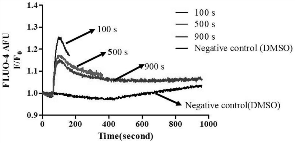
[0041] Example 1 An in vitro detection method for chemical carcinogenicity based on the FPR1 channel, optimization of high-throughput real-time fluorescence detection conditions;
[0042] (1) Cell seed plate
[0043] FPR1-CHO cells in the logarithmic growth phase were divided into 5×10 3 pcs / hole, 1×10 4 pcs / hole, 2×10 4 pcs / hole, 4×10 4 The density of cells / well was planted in 96-well black transparent flat-bottomed cell plates. The next day, the cell confluence was observed under an inverted microscope.
[0044] (2) Dye Incubation
[0045] Add calcium ion dye Fluo-4 AM at a volume of 60 μL per well, and incubate at 37°C for 45min-60min. After incubation, wash 3-4 times with HBSS buffer to remove dye that has not entered the cells. After washing, add HBSS buffer to a final volume of 175 μL.
[0046] (3) Treatment of chemicals to be tested
[0047] The FPR1 agonist fMLP was dissolved in DMSO into a 100 mM sample solution. Dilute with HBSS buffer at a ratio of 1:125, ...
Embodiment 2
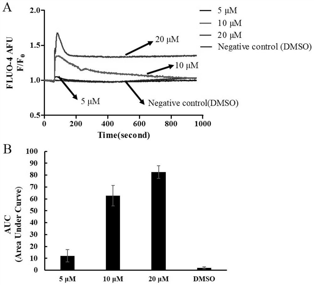
[0056] An in vitro assay of chemical carcinogenicity based on the FPR1 channel, evaluating the carcinogenicity of ochratoxin A (OTA);
[0057] (1) Cell seed plate
[0058] Take FPR1-CHO cells in the logarithmic growth phase in 2×10 4 The density of cells / well was planted in 96-well black transparent flat-bottomed cell plates. On the next day, the degree of cell fusion was observed under a microscope, and the dye incubation could be carried out when it reached more than 70%.
[0059] (2) Dye Incubation
[0060] Add calcium ion dye Fluo-4 AM at a volume of 60 μL per well, and incubate at 37°C for 45min-60min. After incubation, wash 3-4 times with HBSS buffer to remove dye that has not entered the cells. After washing, add HBSS buffer to a final volume of 175 μL.
[0061] (3) Treatment of chemicals to be tested
[0062] OTA was first dissolved in DMSO into 20mM, 10mM, 5mM sample solutions. Dilute them with HBSS buffer at a ratio of 1:125, add 60 μL per well to a 96-well tr...
Embodiment 3
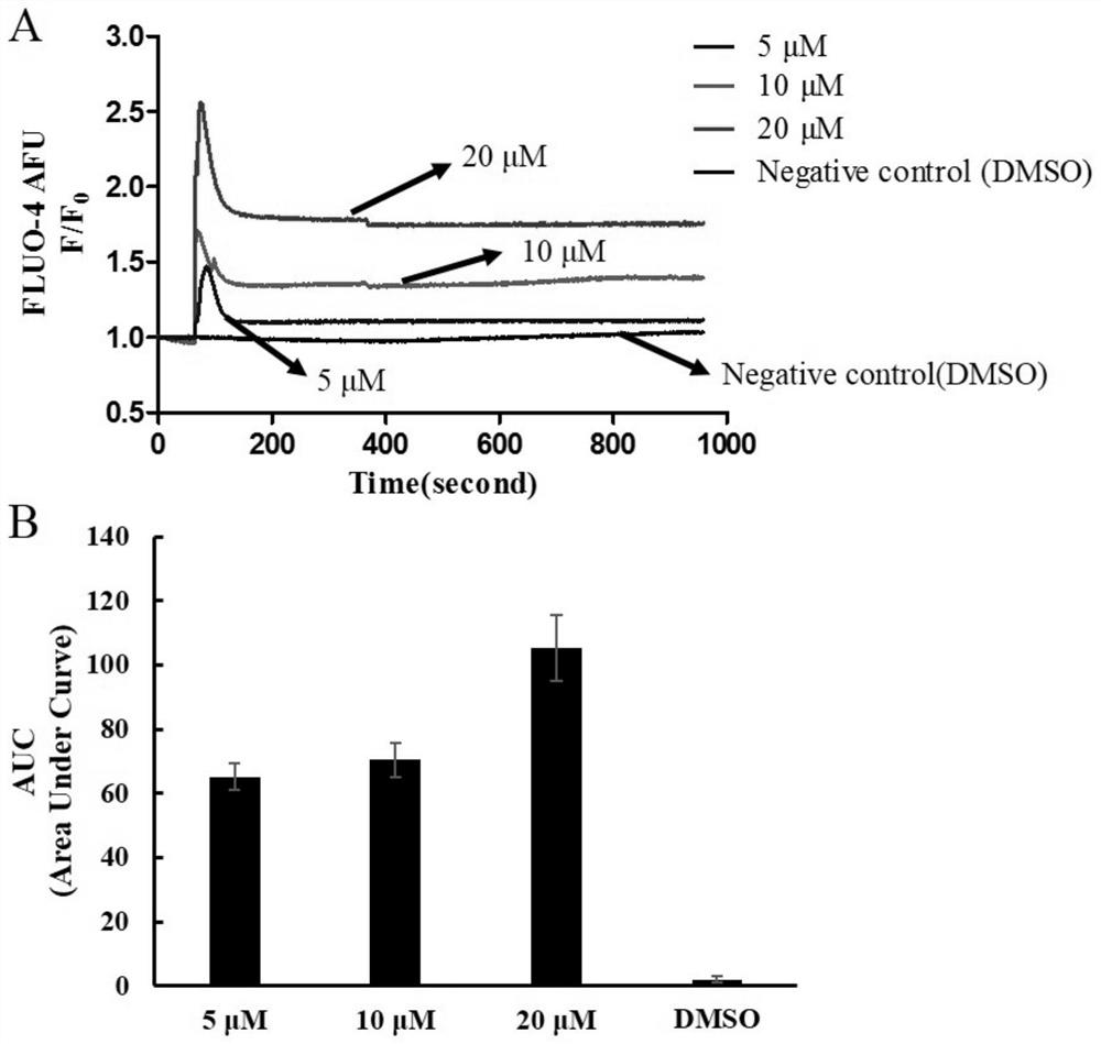
[0069] A chemical carcinogenicity detection method based on FPR1 channel in vitro to evaluate the carcinogenicity of patulin;
[0070] (1) Cell seed plate
[0071] The steps of seeding the cells are the same as in Example 2.
[0072] (2) Dye Incubation
[0073] Dye incubation steps are the same as in Example 2.
[0074] (3) Treatment of chemicals to be tested
[0075] Dissolve patulin in DMSO into sample solutions with concentrations of 20mM, 10mM, and 5mM. Dilute with HBSS buffer at a ratio of 1:125, and add to a 96-well transparent conical-bottom microtiter plate at a volume of 60 μL per well.
[0076] (4) High-throughput real-time fluorescence detection
[0077] The high-throughput real-time fluorescence detection is the same as that in Example 2.
[0078] (5) Results
[0079] Such as image 3 As shown, compared with the negative control, after patulin at the final concentration of 5 μM, 10 μM, and 20 μM acted on FPR1-CHO cells, the intracellular calcium ion concent...
PUM
 Abstract
Description
Claims
Application Information
 - R&D Engineer
- R&D Manager
- IP Professional
- Industry Leading Data Capabilities
- Powerful AI technology
- Patent DNA Extraction
Browse by: Latest US Patents, China's latest patents, Technical Efficacy Thesaurus, Application Domain, Technology Topic, Popular Technical Reports.
© 2024 PatSnap. All rights reserved.Legal|Privacy policy|Modern Slavery Act Transparency Statement|Sitemap|About US| Contact US: help@patsnap.com


