
Endometrial pathological image classification method

An endometrial and pathological image technology, applied in the field of endometrial pathological image classification, achieves the effect of speeding up the reading speed, reducing the heavy workload of screening, and achieving good use effect.

Active Publication Date: 2021-06-11
THE FIRST AFFILIATED HOSPITAL OF MEDICAL COLLEGE OF XIAN JIAOTONG UNIV +1
View PDF4 Cites 2 Cited by
  • Summary
  • Abstract
  • Description
  • Claims
  • Application Information

AI Technical Summary

Problems solved by technology

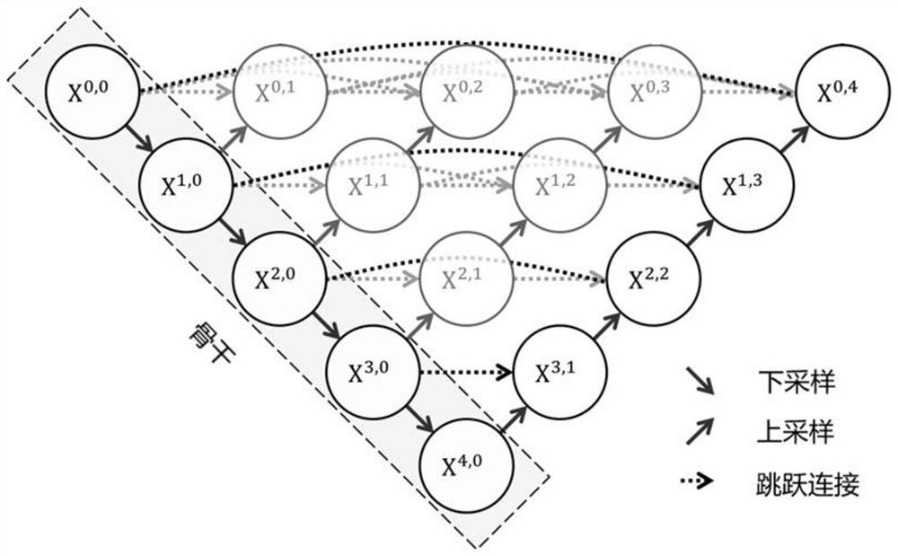
[0003] The technical problem to be solved by the present invention is to provide a method for classifying endometrial pathological images, which needs to quickly identify the endometrium Whether the cells, endometrial cell populations, and endometrial microtissues are pathological, the rapid segmentation of historical endometrial images can be realized by improving the skipping path of the diluted scanner network, and the reading speed can be accelerated, using two LSTM models and Inception-v3 The CNN image model processes image features synchronously, obtains clinical features, and visually, quickly and accurately classifies endometrial cells and microtissue pathology, thereby laying the foundation for the development of endometrial cancer screening and reducing the workload of crowd screening Large size, low work efficiency and other problems, easy to promote and use

Method used


Image

  • Endometrial pathological image classification method
  • Endometrial pathological image classification method

Examples


Embodiment Construction

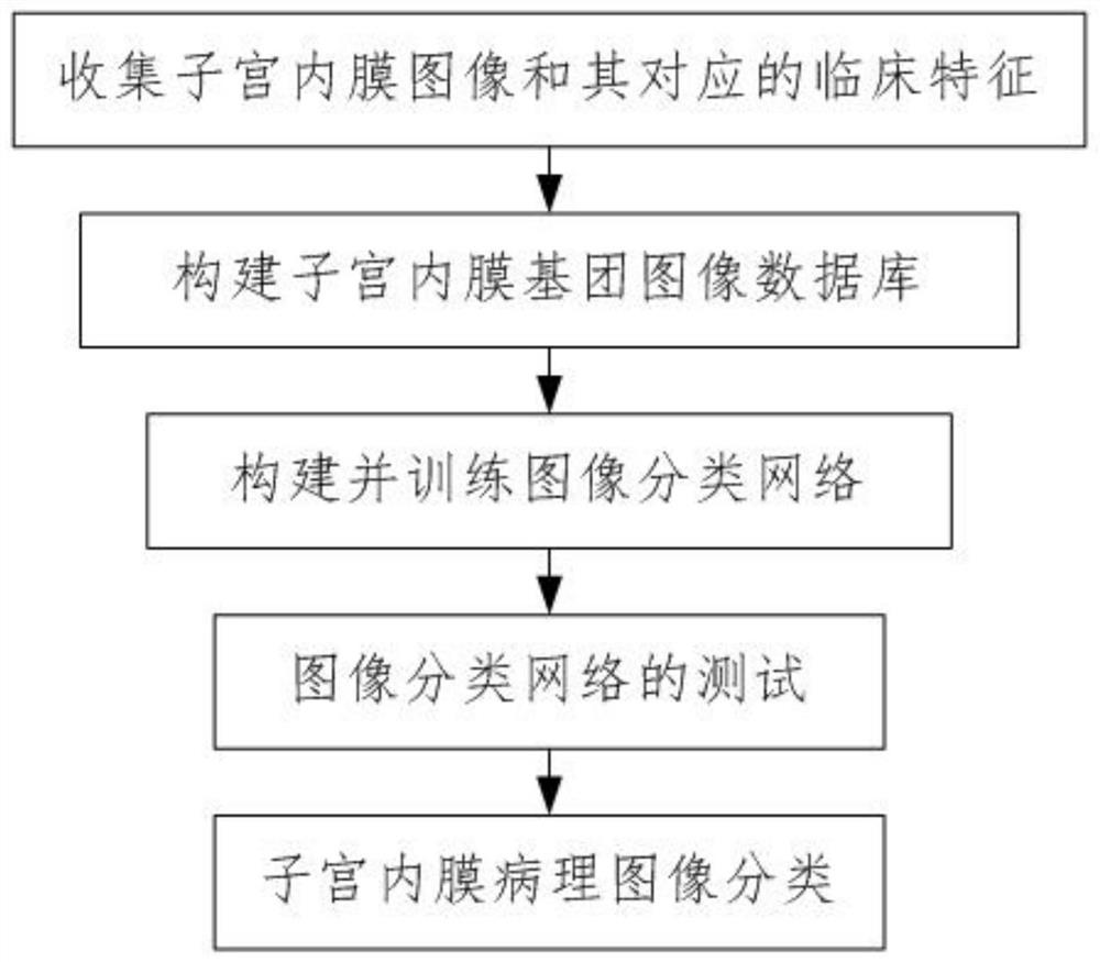
[0042] Such as figure 1 with figure 2 As shown, a subterior pathological image classification method of the present invention includes the following steps:

[0043] Step 1. Collecting the endometrial image and its corresponding clinical features: clinical features corresponding to the historical endometrium image and the endometrial image, the endometrial image including a positive uterine intimal image and a negative uterine intimal image ;

[0044] The clinical features include clinical pathological characteristics, endometrial molecular profiling;

[0045] In this embodiment, the clinical pathological characteristics include the nucleus of the nucleus, and the nucleus is increased, and the nucleus is deeply reduced, and the cells do not need to split.

[0046] The endometrial molecular profile includes a POLE super mutation, MSI high mutation, low copy number, high copy number.

[0047] Step 2, construct the endometrial group image database, the process is as follows:

[0048] ...


PUM


Abstract

The invention discloses an endometrial pathological image classification method. The method comprises the following steps: 1, collecting an endometrial image and a clinical feature corresponding to the endometrial image; 2, constructing an endometrial group image database; 3, constructing and training an image classification network; 4, testing an image classification network; and 5, classifying endometrial pathological images. According to the method, whether endometrial cells, endometrial cell communities and endometrial micro-tissues are diseased or not needs to be rapidly identified, rapid segmentation of historical endometrial images is achieved by improving a dilution scanner network skipping path, the film reading speed is increased, two LSTM models and an Incept ion-v3CNN image model are used for synchronously processing image features, clinical features are obtained. The endometrial cell and micro-tissue pathological classification is vividly, intuitively, quickly and accurately carried out, so that a foundation is laid for the development of endometrial cancer screening work, and the problems of large workload, low working efficiency and the like of crowd screening are solved.

Description

Technical field [0001] The present invention belongs to the field of endometrial pathological image classification, and is specifically related to a uterine intimal pathological image classification method. Background technique [0002] The incidence and mortality rate of endometrial cancer in recent years have an upward trend in the world. In many developing countries, the incidence of endometrial cancer is second in a malignant tumor of female reproductive system. Due to the improvement of living standards, in some developed countries and developed cities such as the United States, the United Kingdom, Japan, and Shanghai, the incidence of endometrial cancer and even more than cervical cancer, which becomes the most common malignant tumor for female reproductive systems. Therefore, the endometrial carcinoma screening will be in the case. Using cytology and micro-woven image to identify endometrial pathological images, however, due to consolidation of hormone levels, ovarian func...

Claims


Application Information

Patent Timeline
IPC IPC(8): G06K9/62G06K9/34G06N3/04G06N3/08G06F16/583G06F16/55G16H10/20G16H30/20
CPCG06N3/08G06F16/583G06F16/55G16H10/20G16H30/20G06V10/267G06N3/045G06N3/044G06F18/24
Inventor 李奇灵钟德星韩露赵蓝波赵惊涛
Owner THE FIRST AFFILIATED HOSPITAL OF MEDICAL COLLEGE OF XIAN JIAOTONG UNIV
Learn More