Lytic vibrio parahaemolyticus bacteriophage RDP-VP-19003 and application thereof
A technology of RDP-VP-19003, Vibrio hemolyticus, applied in the direction of bacteriophage, virus/phage, medical raw materials derived from virus/phage, etc. Effect
- Summary
- Abstract
- Description
- Claims
- Application Information
AI Technical Summary
Problems solved by technology
Method used
Image
Examples
Embodiment 1
[0031] Example 1 Isolation and identification of Vibrio parahaemolyticus BV-19004
[0032] Samples were taken from the diseased sea cucumber lesions, using aseptic operation, streaked on TCBS medium, and cultured at 37°C for 18-24 hours, on the TCBS agar plate appeared round green colonies with neat edges and a diameter of about 2-4. mm; then pick a typical colony and streak it on the Chromagar Vibrio chromogenic medium. After culturing at 37°C for 18-24 hours, purple colonies appear on the Chromagar Vibrio chromogenic medium. Pick typical colonies and continue to streak Line purification was performed 3 times, and then a single colony was picked and inoculated in 5 mL of 2216E liquid medium, and cultured with shaking at 200 rpm at 37°C for 8 hours to obtain a uniform turbid bacterial suspension. Through 16sRNA molecular identification, it was identified as Vibrio parahaemolyticus, and one of them was named BV-19004.
Embodiment 2
[0033] Example 2 Isolation and identification of Vibrio parahaemolyticus phage RDP-VP-19003
[0034] The sewage sample used in the test of the present invention was collected from the surrounding sewage of farmers in Jimo, Qingdao in 2018, and was used as a water sample for the separation of bacteriophage.
[0035] Take 10 ml of sewage, centrifuge at 10,000rpm for 5 minutes, remove larger impurities and most of the bacteria, and then use a 0.22 micron microporous membrane to filter and sterilize. Take 3 milliliters of the filtrate and 3 milliliters of the host bacterial suspension, add them together into 20 milliliters of autoclaved LB broth, and then place them in a 37°C incubator for overnight culture. After culturing, take 5 ml, centrifuge at 10,000 rpm for 5 minutes, and then filter and sterilize with a 0.22 micron microporous filter. The filtrate is the stock solution intended to contain phage. Then, use the double-plate method to identify whether there are phages; pick ...
Embodiment 3
[0037] Example 3 Morphological observation of bacteriophage
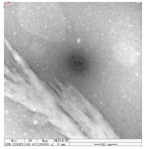
[0038] Drop phage liquid (titer 10 10 pfu / m L) 100 μL, put the membrane side of the copper mesh on the phage droplet, take it off after 10 min, and let it dry naturally in the air for 2-3 min. Then drop a drop of 2% phosphotungstic acid (PTA) aqueous solution on the copper grid for staining, take it off after 10 min, dry it in the air for 10-15 min, observe with an electron microscope, and select a clear phage image to take pictures. Photo by electron microscope ( figure 2 ) It can be seen that the phage has no tail, is a regular hexagon, and the diameter of the head is about 55nm.
PUM
 Abstract
Description
Claims
Application Information
 - Generate Ideas
- Intellectual Property
- Life Sciences
- Materials
- Tech Scout
- Unparalleled Data Quality
- Higher Quality Content
- 60% Fewer Hallucinations
Browse by: Latest US Patents, China's latest patents, Technical Efficacy Thesaurus, Application Domain, Technology Topic, Popular Technical Reports.
© 2025 PatSnap. All rights reserved.Legal|Privacy policy|Modern Slavery Act Transparency Statement|Sitemap|About US| Contact US: help@patsnap.com


