Fluorescent probe for detecting beta-galactosidase as well as preparation method and application of fluorescent probe
A galactosidase and fluorescent probe technology, applied in the preparation of sugar derivatives, biochemical equipment and methods, fluorescence/phosphorescence, etc., can solve the problems of high cost, complex probe synthesis, etc. Good performance and high sensitivity
- Summary
- Abstract
- Description
- Claims
- Application Information
AI Technical Summary
Problems solved by technology
Method used
Image
Examples
Embodiment 1
[0042] A fluorescent probe for detecting β-galactosidase, the preparation method of which comprises the following steps:
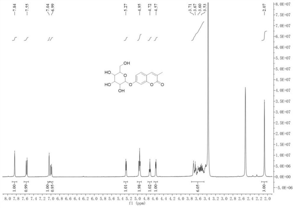
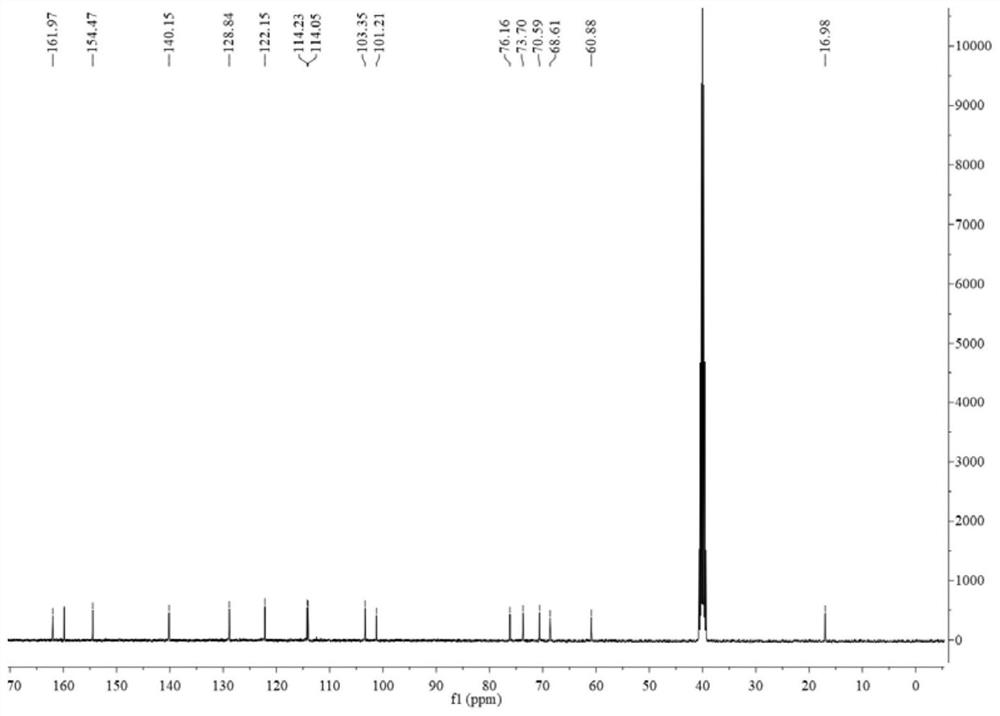
[0043] 1) Synthesis of compound a: Weigh 1g of 2,4-dihydroxybenzaldehyde (7.2mmol), place 1.57g of sodium propionate (16.3mmol) in a 25mL round-bottomed flask, measure 3.16mL of propionic anhydride ( 24.5mmol), 1.15mL of dry triethylamine (8.3mmol) were added to the above system, heated to 170°C and refluxed for 2h, the reaction was complete as monitored by TLC, after the reaction was completed, cooled to room temperature, stirred with water, at this time there were a large amount of white solid separated out, then extracted with ethyl acetate, washed the organic phase three times with water, anhydrous MgSO 4Drying, spin-drying, and then by column chromatography (ethyl acrylate:petroleum ether=1:6, v / v) to obtain a light yellow solid, then recrystallized with ethyl acrylate, after vacuum drying to obtain 1.1g of product a, the yield About 65%. product a ...
Embodiment 2
[0055] Measure the effect of the fluorescent probe obtained in Example 1 and β-galactosidase reaction:
[0056] Add 3mL of fluorescent probe molecules prepared in Example 1 (10 μM, dissolved in PBS buffer solution with a pH value of 7.4 at 37°C) to the cuvette as a control group, test its ultraviolet absorption spectrum and fluorescence spectrum, and then In another cuvette, add 3 mL of fluorescent probe molecules prepared in Example 1 (10 μM, dissolved in PBS buffer solution with a pH value of 7.4) and a mixture of β-galactosidase (0.10 U / mL) as The experimental group tested its ultraviolet absorption spectrum and fluorescence emission spectrum. Such as Figure 7 Shown is the high performance liquid chromatography and mass spectrometry of the fluorescent probe detecting β-galactosidase. It can be seen from the figure that β-galactosidase breaks the D-galactosidic bond and releases the fluorescent group, which illustrates the reaction mechanism of the probe Consistent with a...
Embodiment 3
[0058] Measure the change of the concentration of the fluorescent probe obtained in Example 1 and the β-galactosidase reaction intensity of different concentrations:
[0059] Add 3 mL of fluorescent probe molecules (10 μM, dissolved in PBS buffer solution with a pH value of 7.4 at 37° C.) and different concentrations of β-galactosidase (0 , 0.01, 0.03, 0.05, 0.07, 0.1, 0.15U / mL) mixed solution, after reacting for 15 minutes, test the fluorescence spectrum of each mixed solution. As the concentration of β-galactosidase increases (0-0.10U / mL), the fluorescence intensity increases gradually, and a red shift occurs at the same time. However, when a larger amount of β-galactosidase (0.15U / mL) was added, the fluorescence intensity no longer increased, which indicated that 0.10U / mL β-galactosidase could catalyze the hydrolysis of 10 μM fluorescent probe, different The titration test chart of the concentration of β-galactosidase to the fluorescent probe is as follows Figure 11 show...
PUM
 Abstract
Description
Claims
Application Information
 - R&D Engineer
- R&D Manager
- IP Professional
- Industry Leading Data Capabilities
- Powerful AI technology
- Patent DNA Extraction
Browse by: Latest US Patents, China's latest patents, Technical Efficacy Thesaurus, Application Domain, Technology Topic, Popular Technical Reports.
© 2024 PatSnap. All rights reserved.Legal|Privacy policy|Modern Slavery Act Transparency Statement|Sitemap|About US| Contact US: help@patsnap.com


