Fluorescent probe for detecting cell hydroxyl radical, and synthesis method and use
A fluorescent probe and free radical technology, applied in the field of biological detection technology and clinical medical detection, to achieve the effect of good permeability, good selectivity and short response time
- Summary
- Abstract
- Description
- Claims
- Application Information
AI Technical Summary
Problems solved by technology
Method used
Image
Examples
Embodiment 1
[0038] Probe Synthesis
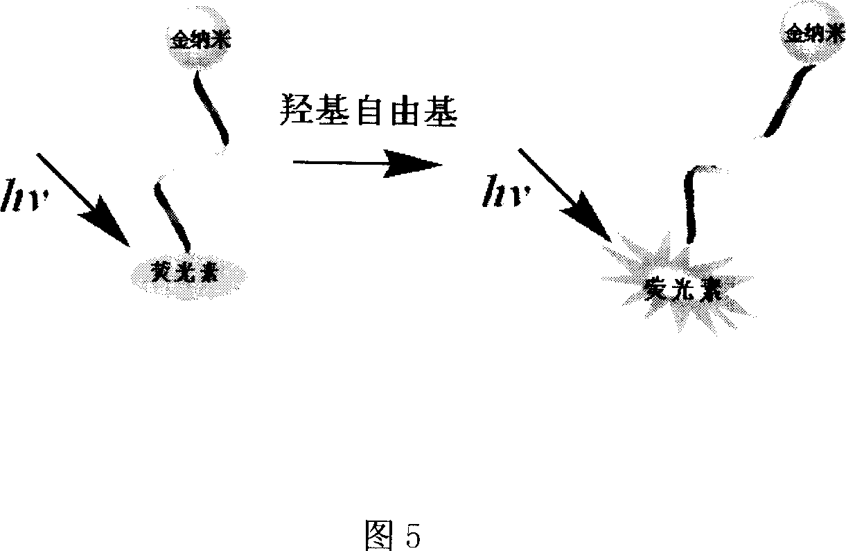
[0039]
[0040] In the formula
[0041] AGGGTTAGGG: DNA oligonucleotide chain single strand, any base sequence, the number of bases is 2-30;
[0042] Fluorescein: X 1 , X 2 = F, Cl, CH 3 , OCH 3 ;
[0043] SH: Mercapto
[0044] Gold nanoparticles: 3-32nm in diameter.
[0045] a. Take 150.0 mL of chloroauric acid solution with a weight concentration of 0.01% in a 250 mL Erlenmeyer flask, heat to reflux, add 5.0 mL of trisodium citrate solution with a weight concentration of 2.0% under stirring, and continue to reflux for 20 minutes. placed under cooling to obtain a dark red gold nanoparticle solution; the transmission electron microscope results showed that the average particle diameter of the particles was 15.5nm, and the particle concentration was 1.2×10 according to the complete reaction estimation. 15 / L.
[0046] b. Mix the gold nanoparticle solution with the 5' end-modified fluorescein and the 3' end-modified thiol-modified DNA single ...
Embodiment 2
[0048] a. Take 150.0 mL of chloroauric acid solution with a weight concentration of 0.02% in a 250 mL conical flask, heat to reflux, add 7.0 mL of trisodium citrate solution with a weight concentration of 1.0% under stirring, and continue to reflux for 10 minutes. Place it under cooling to obtain a deep red gold nanoparticle solution; the transmission electron microscope results show that the average particle diameter of the particles is 15.9nm, and the particle concentration is 1.2×10 according to the complete reaction estimation. 15 / L.
[0049] b. Mix the gold nanoparticle solution with the 5'-end modified fluorescein and the 3'-end sulfhydryl-modified DNA single-strand according to the ratio of 1:500, leave it at room temperature for 12 hours, and then adjust the pH to 7.8 with phosphate buffer ;Then the sodium chloride solution that is 0.5mol / L is mixed with the sodium chloride solution whose concentration is 0.5mol / L through adjusting the pH value, and is placed at room ...
Embodiment 3
[0051] a. Take 150.0 mL of chloroauric acid solution with a weight concentration of 0.015% in a 250 mL Erlenmeyer flask, heat to reflux, add 6.0 mL of trisodium citrate solution with a weight concentration of 1.5% under stirring, and continue to reflux for 15 minutes. placed under cooling to obtain a dark red gold nanoparticle solution; the transmission electron microscope results showed that the average particle diameter of the particles was 15.7nm, and the particle concentration was 1.2×10 according to the complete reaction estimation. 15 / L.
[0052] b. Mix the gold nanoparticle solution with the 5'-end modified fluorescein and the 3'-end sulfhydryl-modified DNA single-strand according to the ratio of 1:350, leave it at room temperature for 18 hours, and then adjust the pH to 7.5 with phosphate buffer ; then the mixed solution through adjusting the pH value and the sodium chloride solution whose concentration is 3mol / L are mixed evenly by 1: 0.2 parts by volume, and placed ...
PUM
| Property | Measurement | Unit |
|---|---|---|
| Diameter | aaaaa | aaaaa |
Abstract
Description
Claims
Application Information
 - R&D
- Intellectual Property
- Life Sciences
- Materials
- Tech Scout
- Unparalleled Data Quality
- Higher Quality Content
- 60% Fewer Hallucinations
Browse by: Latest US Patents, China's latest patents, Technical Efficacy Thesaurus, Application Domain, Technology Topic, Popular Technical Reports.
© 2025 PatSnap. All rights reserved.Legal|Privacy policy|Modern Slavery Act Transparency Statement|Sitemap|About US| Contact US: help@patsnap.com


