
Prothrombin time detection test strip and preparation method thereof

A technology of prothrombin time and test strips, which is applied in the field of detection and can solve the problems of expensive instruments, complicated operations, and high costs

Pending Publication Date: 2020-05-19
杭州联晟生物科技有限公司
View PDF7 Cites 1 Cited by
  • Summary
  • Abstract
  • Description
  • Claims
  • Application Information

AI Technical Summary

Problems solved by technology

Its main disadvantages are complex and professional operation, relatively high cost, and expensive instruments, which are not suitable for testing in ambulances or outdoors.
[0005] Although individual patents describe the use of test cards and supporting instruments for testing, such as CN 106680339A mentions the use of electrochemical methods to detect prothrombin, but due to the use of two holes and two reagents, the test results are due to material problems or other process problems It is difficult to achieve process, and the interference of cells with too high or too low volume cannot be ruled out, which reduces the accuracy of test results

Method used


Image

  • Prothrombin time detection test strip and preparation method thereof
  • Prothrombin time detection test strip and preparation method thereof
  • Prothrombin time detection test strip and preparation method thereof

Examples


preparation example Construction

[0043] A preparation method of a prothrombin time detection test strip, comprising the steps of:

[0044] Step 1, the preparation of reaction reagent;

[0045] Prepare materials according to the following formula: the formula includes: 4-6 parts of tissue factor, 0.4-0.5 parts of phosphatidylcholine, 0.1-5 parts of film-forming agent, 2-12 parts of stabilizer, 0.1-1 part of preservative , 0.5-1 parts of surfactant, 2-5 parts of coagulant;

[0046] Add tissue factor and phosphatidylcholine into the buffer and mix, then magnetically stir to disperse, then add film forming agent, stabilizer, preservative, surfactant, coagulant, use 100ml concentration of 0.1-1M, pH value is 6 -8 buffer is stirred, stirred for 2 hours, and stored in the refrigerator for use; as an example, the buffer includes: phosphate buffer, TRIS buffer, citrate buffer, MES buffer.

[0047] Step 2, soak the carrier film in the reaction reagent for 5 minutes; after drying, the reagent layer 400 is obtained, an...

Embodiment 1

[0051] Remove the erythrocyte separation layer 500

[0052] 1. Preparation of reagent layer 400 solution: Add 5g of rabbit brain powder and 0.5g of phosphatidylcholine into the buffer and mix, then magnetically stir to disperse, then add 2g of hydroxyethyl cellulose, 2g of bovine serum albumin, 1g Trehalose, 1g of polyethylene glycol 6000, and 0.1g of Proclin300, 0.5g of triton X-405, 2g of calcium chloride, 100ml concentration of 0.5M, pH value of 7.2 phosphate buffer after stirring for 2 hours , kept in the refrigerator for later use.

[0053] 2. Soak the glass fiber membrane in the above solution for 5 minutes, and dry it in a 45-degree oven for 30 minutes to form the reagent layer 400.

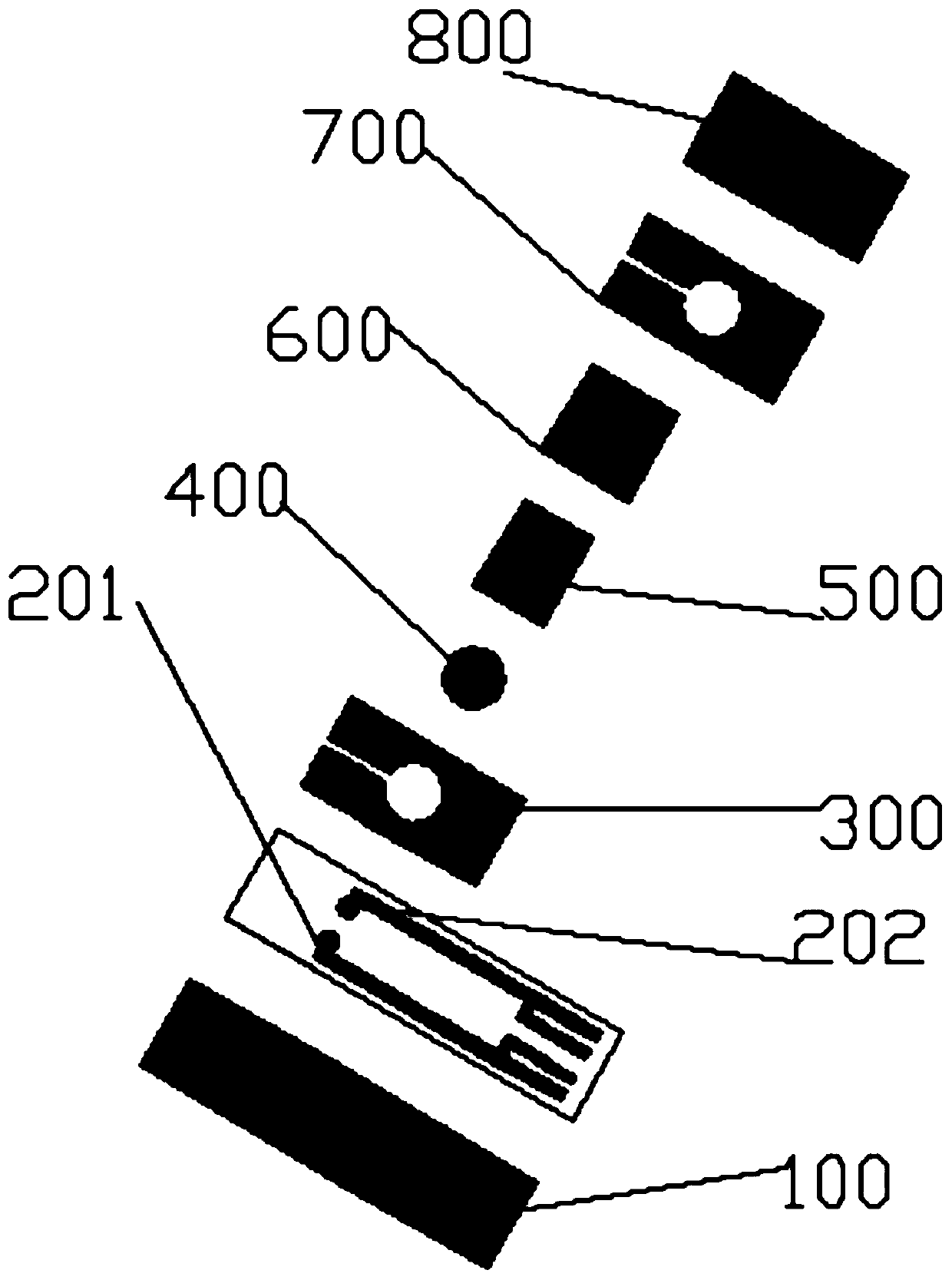
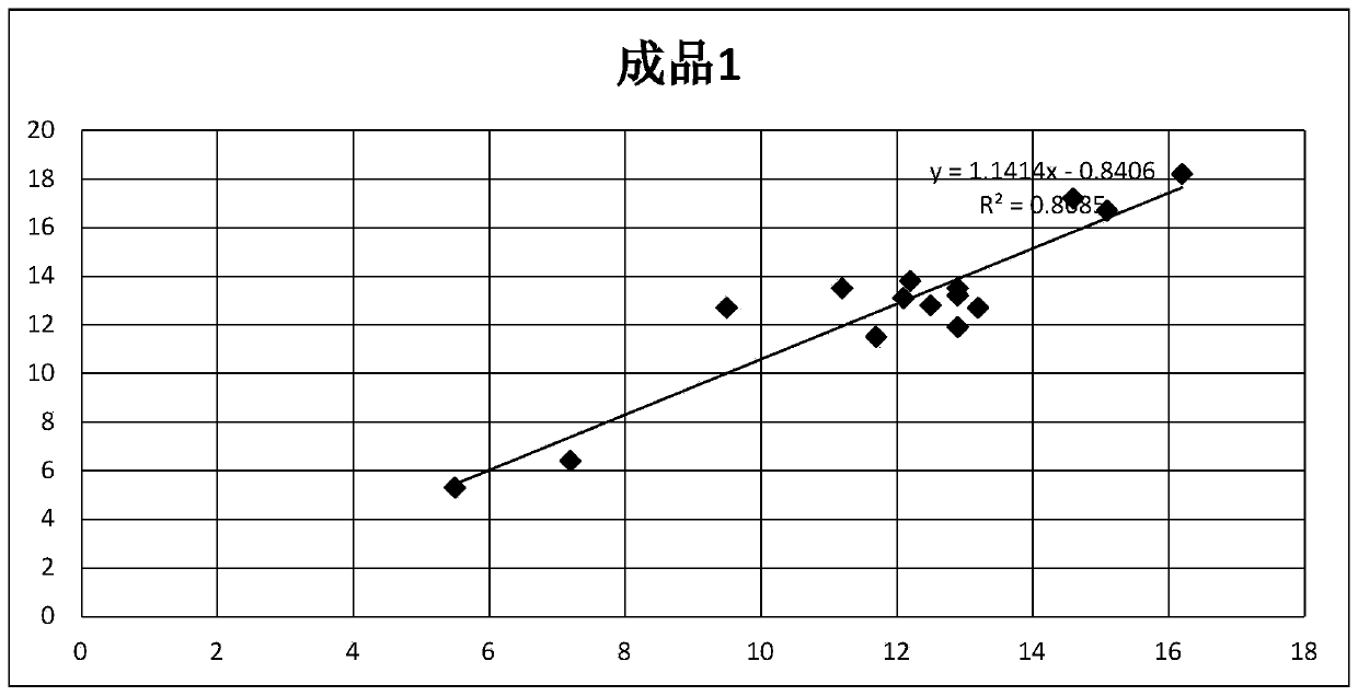
[0054] 3. Combine the prepared electrode 200, double-sided adhesive tape 300, reagent layer 400, diffusion membrane 600, and hydrophilic membrane 700 into a finished product 1.

Embodiment 2

[0056] Compared with embodiment 1, the erythrocyte separation layer 500 is increased

[0057] 1. Preparation of reagent layer 400 solution: Add 5g of rabbit brain powder and 0.5g of phosphatidylcholine into the buffer and mix, then magnetically stir to disperse, then add 2g of hydroxyethyl cellulose, 2g of bovine serum albumin, 1g Trehalose, 1g of polyethylene glycol 6000, and 0.1g of Proclin300, 0.5g of triton X-405, 2g of calcium chloride, 100ml concentration of 0.5M, pH value of 7.2 phosphate buffer after stirring for 2 hours , kept in the refrigerator for later use.

[0058] 2. Soak the glass fiber membrane in the above solution for 5 minutes, and dry it in a 45-degree oven for 30 minutes to form the reagent layer 400.

[0059] 3. Follow the method of 1 to assemble the prepared electrode 200, double-sided adhesive tape 300, reagent layer 400, erythrocyte separation layer 500, diffusion membrane 600, and hydrophilic membrane 700 to form the finished product 2 in Figure 2. ...


PUM


Abstract

The invention discloses a prothrombin time detection test strip and a preparation method thereof. The test strip comprises a hydrophilic layer, a diffusion layer arranged below the hydrophilic layer,a first bonding layer arranged between the hydrophilic layer and the diffusion layer, a red blood cell separation layer arranged below the diffusion layer, a reagent layer arranged below the red bloodcell separation layer, an electrode layer arranged below the reagent layer, a substrate arranged below the electrode layer, and a second bonding layer which fixes the reagent layer and is located between the first bonding layer and the substrate. The detection test strip is advantaged in that a single-hole single-reagent method is adopted to test the prothrombin time, the test strip can be suitable for testing samples with different hematocrit, is matched with a dry-type multifunctional analyzer for use, and is rapid in detection, and the obtained detection result has high accuracy.

Description

technical field [0001] The invention relates to the detection field, in particular to a prothrombin time detection test strip and a preparation method thereof. Background technique [0002] Blood coagulation is a relatively common inspection item, especially some patients with thrombotic diseases need to do blood coagulation inspection, this inspection item is also necessary before surgery, and some diseases also require patients to do blood coagulation before oral anticoagulant drugs check. The blood coagulation examination is very useful. The results of the examination can promptly know whether the patient has some hemostatic dysfunction, so that various possible emergency measures can be taken before the operation, and it also has the effect of preventing surgical bleeding. [0003] The blood coagulation item belongs to one of the clinical inspection items of the laboratory department, and belongs to the examination of thrombotic diseases. It is a must-check item before...

Claims


Application Information

Patent Timeline
IPC IPC(8): G01N27/26G01N27/30G01N27/333
CPCG01N27/26G01N27/30G01N27/333
Inventor 陈军刚
Owner 杭州联晟生物科技有限公司
Learn More