
Surface-modified microorganism and preparation method and application thereof

A surface modification and microbial technology, applied in the biological field, can solve the problems of unfavorable expansion of production, inability to improve the effect of bacterial prevention and treatment, and short residence time, so as to improve survival rate and tolerance, increase colonization and enrichment ability, The effect of reducing toxic side effects

Pending Publication Date: 2020-01-21
RENJI HOSPITAL AFFILIATED TO SHANGHAI JIAO TONG UNIV SCHOOL OF MEDICINE
View PDF7 Cites 7 Cited by
  • Summary
  • Abstract
  • Description
  • Claims
  • Application Information

AI Technical Summary

Problems solved by technology

Although these methods have improved the oral efficiency of oral probiotics to varying degrees, they still cannot improve the preventive and therapeutic effects of these bacteria in the body. For example, the complex gastrointestinal environment (including intestinal microorganisms, drugs, diet, strong acid, Insufficient survival of probiotics caused by bile acids, etc.), and the inhibition of probiotic colonization in the intestinal tract by the package, resulting in a short residence time in the intestinal tract
In addition, cumbersome preparation methods and harsh separation procedures are not conducive to industrial expansion of production

Method used


Image

  • Surface-modified microorganism and preparation method and application thereof
  • Surface-modified microorganism and preparation method and application thereof
  • Surface-modified microorganism and preparation method and application thereof

Examples


specific Embodiment

[0105] The experimental methods used in the examples are conventional research methods in the fields of biochemistry, molecular biology or medical detection. Wherein, Escherichia coli Nissle 1917 (hereinafter referred to as EcN), Enterococcus faecalis (E.faecalis), Staphylococcus aureus (S.aureus) and Salmonella typhimurium (Salmonella typhimurium) used in the present invention were purchased from China General Microorganism Culture Collection center.

[0106] Cell lines (Hela, CT26 and 4T1) were from the American Species Culture Collection (ATCC).

[0107] Plasmids pBBR1MCS2-Tac-mCherry, pBBR1MCS2-Tac-GFP and pMD18-luxCDABE were purchased from domestic suppliers.

[0108] Bacteria surrounded by cell membranes

Embodiment 1

[0110] EcN (E. coli Nissle) carrying pBBR1MCS2-Tac-mCherry, pBBR1MCS2-Tac-GFP or pMD18-luxCDABE were grown overnight at 37°C in LB medium containing antibiotics. E. faecalis, S. aureus and S. typhimurium were grown in tryptic soy broth (TSB) medium at 37°C. The overnight culture was diluted 1:50 into fresh LB medium and grown at 37 °C for 3 h. Bacteria were harvested by centrifugation at 4200 rpm for 10 minutes and resuspended in ice-cold PBS. Bacterial counts were determined by diluting the bacterial suspension, growing overnight at 37°C on LB agar plates and counting colony forming units (CFU).

Embodiment 2

[0112] Red blood cell membranes were prepared according to the method reported in reference 1. Briefly, whole blood was extracted through the eye socket from anesthetized male ICR mice (6–8 weeks) purchased from Jiesijie Experimental Animal Center (Shanghai, China), and then centrifuged at 800 g for 5 min at 4 °C to Remove serum and buffy coat. After three washes in cold 1 × PBS, the obtained packaged RBCs were suspended in 0.25 × PBS on ice for 30 min and then centrifuged at 10000 rpm for 5 min to remove hemoglobin. The pink pellet was washed with cold 1x PBS and stored at -20 °C for further use. The resulting cell membranes are shown in Figure 2B, where Figure 2A shows (untreated) erythrocytes.

[0113] All animal procedures complied with the Shanghai Medical Laboratory Animal Care Guidelines. The animal protocol was approved by the Animal Care and Use Committee of Shanghai Jiao Tong University School of Medicine.


PUM


Abstract

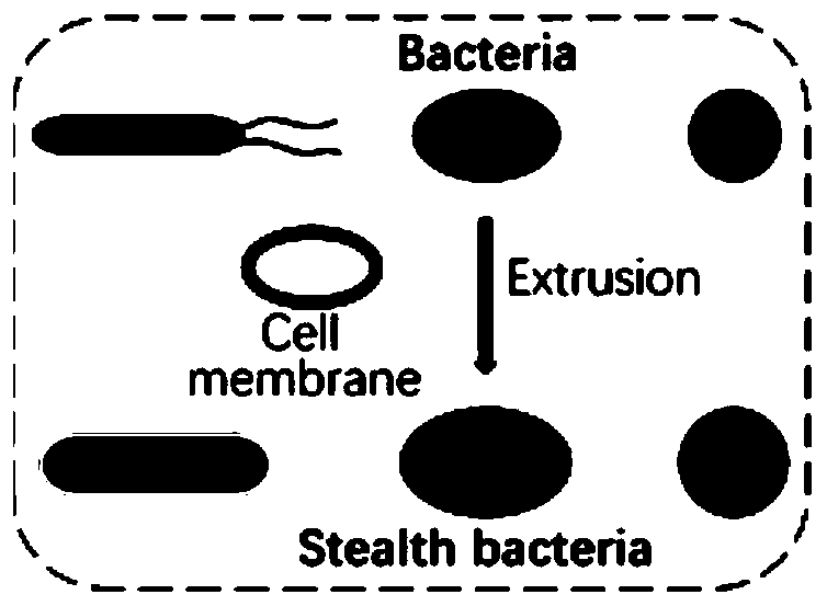
The invention belongs to the field of biotechnologies, and relates to a microorganism wrapped by biological films, and the microorganism is bacteria or fungus; and the biological films are used for blocking surface immunogenicity or other toxic and side effect substances.

Description

technical field [0001] This application belongs to the field of biotechnology, and relates to a method for modifying the surface of microorganisms using biofilms, prepared bacteria and applications, specifically, to a method for modifying the surfaces of microorganisms using biofilms, surface-modified microorganisms prepared by this method, and its application. Background technique [0002] Bacteria are ubiquitous microorganisms that play an important role in maintaining ecological stability, especially in maintaining homeostasis and health in mammals. Due to the unique advantages of bacteria, such as gene editing, rapid reproduction, and easy enrichment in disease sites, especially tumor sites, bacteria have the potential to become biological imaging, diagnostic and therapeutic tools or drugs. [0003] At present, many skilled in the art have invested a lot of research in engineering oral probiotics and bacteria-mediated disease treatment, but the application of bacteria i...

Claims


Application Information

Patent Timeline
Patent Type & Authority Applications(China)
IPC IPC(8): A61K9/50A61K47/46A61K35/742A61K35/747A61K35/745A61K35/741A61P1/00A61P29/00A61P35/00A61K49/00C12N1/20C12N1/16C12R1/19C12R1/01C12R1/445C12R1/07C12R1/145C12R1/225C12R1/125C12R1/645
CPCA61K9/5068A61K35/742A61K35/747A61K35/745A61K35/741A61P1/00A61P29/00A61P35/00A61K49/005C12N1/20C12N1/16A61K2300/00
Inventor 刘尽尧庞燕曹浈萍王馨悦
Owner RENJI HOSPITAL AFFILIATED TO SHANGHAI JIAO TONG UNIV SCHOOL OF MEDICINE
Learn More