Preparation method of decellularized nerve scaffold and effect evaluation method of preparation method

An evaluation method and decellularization technology, applied in the field of nerve scaffold materials, can solve the problems of inability to fully reflect the decellularization effect of scaffolds and the limitations of detection methods

Inactive Publication Date: 2019-11-19
BINZHOU MEDICAL COLLEGE
View PDF4 Cites 2 Cited by
  • Summary
  • Abstract
  • Description
  • Claims
  • Application Information

AI Technical Summary

Problems solved by technology

[0009] At present, the commonly used evaluation methods for the decellularization effect of decellularized neural scaffolds include cytotoxicity, collagenase sensitivity in vitro, cytocompatibility, and local reaction after implantation. These detection methods are limited and cannot fully reflect the decellularization of scaffolds. Effect

Method used


Image

  • Preparation method of decellularized nerve scaffold and effect evaluation method of preparation method
  • Preparation method of decellularized nerve scaffold and effect evaluation method of preparation method
  • Preparation method of decellularized nerve scaffold and effect evaluation method of preparation method

Examples


Embodiment 1

[0102] Example 1: Preparation of decellularized neural scaffold

[0103] The invention adopts the composite method of TritonX-100 plus freeze-thaw enzyme to prepare the decellularized nerve scaffold.

[0104] Experimental animals: healthy adult SD rats, male or female, weighing 180-200 g, provided by Jinan Pengyue Experimental Animal Breeding Co., Ltd.

[0105] Main equipment: -80 refrigerator (Haier brand), rotary constant temperature oscillator (produced by Shanghai Julai Experimental Instrument Co., Ltd.), low temperature shaker (produced by Shanghai Jiechen Instrument Co., Ltd.), cryostat (Leica brand).

[0106] Main reagents: Triton X-100, penicillin G, streptomycin (not mentioned in the following specific steps, please add), trypsin (Gibco), liquid nitrogen, DNase-I, RNase-A (sigma), PBS buffer Liquid (produced by Beijing Zhongshan Jinqiao Biotechnology Co., Ltd.), OCT embedding agent (Lycra).

[0107] Specific steps: weigh the rats, anesthetize the SD rats with 3.5% c...

Embodiment 2

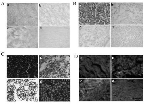
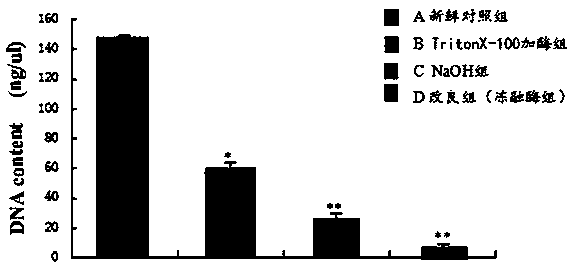
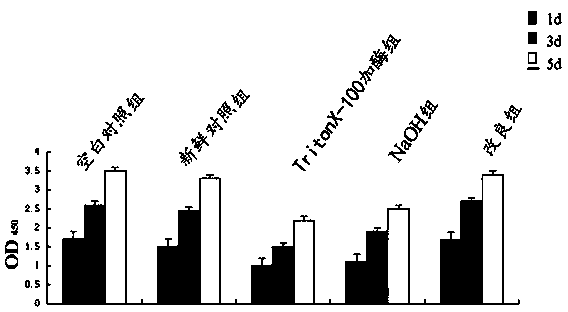
[0110] Example 2: Evaluation of the preparation effect of decellularized neural scaffolds

[0111] Group A (untreated group): fresh control nerves;

[0112] Group B (TritonX-100 enzyme group): decellularized nerve scaffolds;

[0113] Group C (NaOH group): decellularized nerve scaffolds;

[0114] Group D (TritonX-100 plus freeze-thaw enzyme group): decellularized nerve scaffolds prepared by the above preparation method;

[0115] 2.1 Histomorphological observation of decellularized nerve scaffolds

[0116] The fresh control nerves of group A (untreated group) were stained: fixed with 4% paraformaldehyde (4% paraformaldehyde, PFA) and 3% glutaraldehyde, 4°C overnight, sucrose gradient Dehydrate (1 day with 15% sucrose, 2-3 days with 30% sucrose), freeze in liquid nitrogen and use OCT embedding agent for OCT embedding. Tissues were sliced ​​(10 μm) by a cryostat. After the frozen sections were mounted, they were placed at room temperature to dry naturally, stored at -20 degree...


PUM


Abstract

The invention discloses a preparation method of a decellularized nerve scaffold and an effect evaluation method of the preparation method. According to the preparation method, the decellularized nervescaffold is prepared by adopting a mode of combining TritonX-100 and a freeze-thaw enzyme. The decellularized effect on the decellularized nerve scaffold is evaluated through ways of morphological observation, ultrastructure observation, cytotoxicity detection, biocompatibility detection, complex frozen section observation and the like. The result shows that the decellularized nerve scaffold which is prepared by adopting a mode of combining the TritonX-100 and the freeze-thaw enzyme has the advantages of sufficient decellularized effect, complete three-dimensional space structure, smooth canals, good compatibility with cells and tissues, slight inflammatory reaction and good cell growth and proliferation state, is suitable for adhesion growth of cells, and is an ideal scaffold material.

Description

Technical field: [0001] The invention relates to a preparation method of a decellularized nerve scaffold and an effect evaluation method thereof, belonging to the technical field of nerve scaffold materials. Background technique: [0002] Spinal cord injury is a common disease with a high disability rate, which not only endangers the life of patients, but also reduces the quality of life of patients. The huge medical expenses bring a heavy economic burden to families and the medical and health system. Therefore, it is extremely urgent to find effective therapeutic measures for spinal cord injury. With the development of tissue engineering, tissue engineering has become a research boom in the treatment of repairing central nervous system injuries. The key is to apply ideal scaffold materials, select suitable seed cells, combine with signal factors that promote cell growth, and construct tissue engineering complexes. . Scaffolds are frame materials that support the growth of...

Claims


Application Information

Patent Timeline
IPC IPC(8): A61L27/36A61L27/50C12Q1/68C12Q1/02
CPCA61L27/3604A61L27/3675A61L27/3687A61L27/3691A61L27/50A61L2430/32A61L2430/40C12Q1/68G01N33/5005
Inventor 黄飞张璐萍张春雷周帅冯国营田婷高李敏李希凯
Owner BINZHOU MEDICAL COLLEGE
Why Eureka
Try Eureka