
EV71 single-stranded DNA aptamer and chemiluminescent detection kit for detecting enterovirus type-71 by utilizing double aptamers

A technology of aptamer and kit, applied in the field of qualitative detection of EV71 virus, to achieve high specificity, simple and fast detection operation

Active Publication Date: 2019-05-10
ZHENJIANG NO 1 PEOPLES HOSPITAL
View PDF4 Cites 2 Cited by
  • Summary
  • Abstract
  • Description
  • Claims
  • Application Information

AI Technical Summary

Problems solved by technology

However, there is no relevant report on the detection of EV71 based on aptamer technology.

Method used


Image

  • EV71 single-stranded DNA aptamer and chemiluminescent detection kit for detecting enterovirus type-71 by utilizing double aptamers
  • EV71 single-stranded DNA aptamer and chemiluminescent detection kit for detecting enterovirus type-71 by utilizing double aptamers
  • EV71 single-stranded DNA aptamer and chemiluminescent detection kit for detecting enterovirus type-71 by utilizing double aptamers

Examples


Embodiment 1

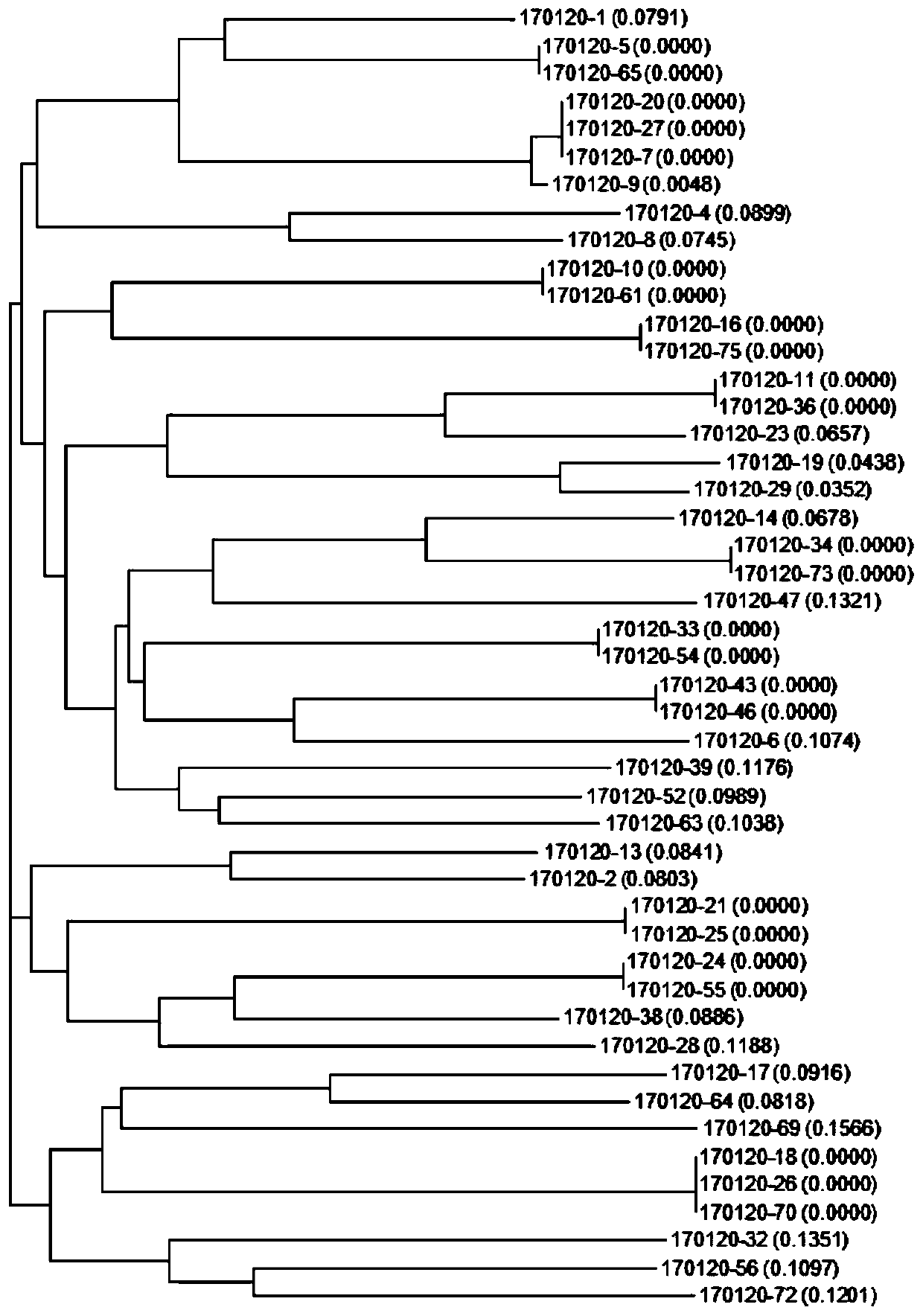
[0035] Screening of EV71 aptamers:

[0036] 1) Main reagents:

[0037] Binding Buffer: 1*PBS, 5mM MgCl 2 , 1% BSA, 1μg / ml tRNA, 0.02% tween-20, 5mM imidazole Washing Buffer: 1*PBS, 5mM MgCl 2 , 0.02% tween-20

[0038] Table 1 Screening library and primer sequences

[0039]

[0040] 2) The first round of SELEX

[0041] a.library was dissolved in water to a final concentration of 20 μM.

[0042] b. Configuration extension system

[0043]

[0044] The extension program is as follows: 95°C for 60s, 58°C for 60s, 72°C for 60min

[0045] c. Add the product to SA agarose bead and incubate at room temperature for 10 minutes to wash away unbound sequences.

[0046] d. Add 150mM NaOH and incubate at room temperature for 5min, aspirate the supernatant, and add 300mM HCl to neutralize.

[0047] e. Using water as a control, Nanodrop measures the concentration of modified single-stranded DNA.

[0048] f. Denaturation at 95°C for 5 minutes, add Binding Buffer to 1ml after ice ...

Embodiment 2

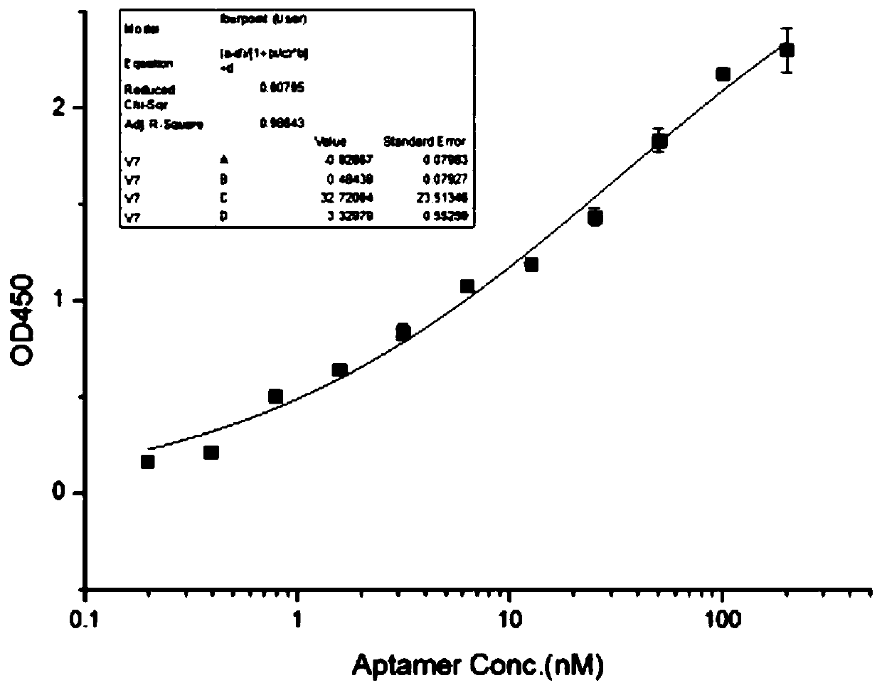
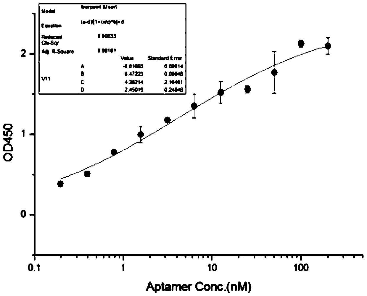
[0082] Aptamer affinity assay (ELISA):

[0083] a. Dilute the protein with PBS to a final concentration of 200ng / ml, set 2 replicate wells for each experimental group, add 50μl to each well. After covering the plate with sealing film, keep overnight at 4°C.

[0084] b. Washing: fill each well with Washing Buffer, let it stand for 5 seconds, pour off the washing solution, and repeat 3 times. Pat dry one last time.

[0085] c. Add 150 μl of PBS containing 1% BSA to each well to block, and incubate at room temperature for 1 hour.

[0086] d. Plate washing: the operation is the same as step b.

[0087] e. Dilute aptamer to the target concentration with Binding Buffer.

[0088] f. Take the coated strip and add 50 μl to each well. Incubate at room temperature for 1h.

[0089] g. Plate washing: the operation is the same as step b.

[0090] h. Dilute Streptavidin-HRP with Aassay Diluent 1:200. Add 50 μl to each well. Incubate at room temperature for 0.5h.

[0091] i. Washing...

Embodiment 3

[0096] Aptamer Specificity Assay (ELISA):

[0097] Referring to the experimental method in Example 2, four coating concentrations of 100ng / ml, 200ng / ml, 500ng / ml, and 1000ng / ml were set up, and the amount of aptamer fixed in each well was 200nM. The result is as Figure 5 As shown, the BSA protein at each coating concentration did not bind to the aptamer. Under the condition of higher coating concentration, the aptamer can also bind to Coxsackie virus A16 (cox) to a certain extent, but the binding ability is significantly lower than that of enterovirus 71 (EV).


PUM


Abstract

The invention relates to an EV71 single-stranded DNA aptamer and a chemiluminescent detection kit for detecting an enterovirus type-71 by utilizing double aptamers. The aptamer is an aptamer V11 having a sequence shown as SEQ ID NO.1, an aptamer V21 having a sequence shown as SEQ ID NO.2, and an aptamer V7 having a sequence shown as SEQ ID NO.3. The kit takes the aptamer V21 as a capture probe andthe aptamer V11 as a signal probe. The chemiluminescent detection kit specifically comprises capture probe coupled immunomagnetic beads, an assay buffer solution, a signal probe coupled enzyme conjugate, an SA-HRP solution and a luminescent substrate solution. The kit has high specificity for EV71 detection and can qualitatively detect EV71 from a cerebrospinal fluid, a herpes fluid, a pharyngealswab, an anal swab and other samples of a suspected hand-foot-mouth disease patient. By utilizing the kit, detection operation is simple and rapid, and the detection process only needs about 45 minutes.

Description

technical field [0001] The invention relates to a single-stranded DNA aptamer for EV71, and a chemiluminescent kit for detecting enterovirus 71 (EV71) using a double aptamer and an application thereof, which is suitable for qualitative detection of EV71 virus. Background technique [0002] Hand, foot and mouth disease (HFMD) is an infectious disease caused by enterovirus, which has occurred sporadically in various places in recent years. The disease is common in preschool children, especially infants, and can cause fever, rashes, and ulcers on the hands, feet, and mouth. Generally, the prognosis is good. Individual patients can cause complications such as myocarditis, pulmonary edema, and aseptic meningoencephalitis. disease. The source of infection of hand, foot and mouth disease is patients and latent infections, mainly through close contact with the virus in the feces, oral secretions, and skin herpes fluid of infected persons, through the fecal-oral route or the respira...

Claims


Application Information

Patent Timeline
Patent Type & Authority Applications(China)
IPC IPC(8): C12N15/115G01N33/58G01N33/543
Inventor 茅凌翔邹欣然吴静顾佳奇贡佳怡沈俐
Owner ZHENJIANG NO 1 PEOPLES HOSPITAL
Learn More