Lentiviral expression vector for permanently labeling nuclei and labeling method
An expression vector and lentivirus technology, applied in the field of biomedicine, can solve the problems of inconvenient living cell division, motion observation, research, inability to achieve permanent nuclear labeling, easy quenching of fluorescent signals, etc., and achieve good application prospects and development space, Wide application and wide range of effects
- Summary
- Abstract
- Description
- Claims
- Application Information
AI Technical Summary
Problems solved by technology
Method used
Image
Examples
Embodiment 1
[0043] Embodiment 1 lentiviral expression vector construction
[0044] Design the primers of the nucleotide sequence of EGFP, and add the nuclear localization signal (NLS) sequence shown in SEQ ID NO:4 at the 5' end of the upstream primer, and send it to Shanghai Yingjun Company for synthesis, and the sequence of the upstream primer is as SEQ ID Shown in NO:6, the downstream primer sequence is shown in SEQ ID NO:7. Use the PEGFP-C2 vector as a template to carry out PCR amplification. After the product is subjected to 1% agarose electrophoresis, the target band is recovered and connected to the PCDH-CMV-MCS-EF1-RFP vector treated with XbaI and EcoRI. After transformation Positive clones were extracted and sequenced. The sequencing results were consistent with those shown in SEQ ID NO:5, and the plasmids were extracted. That is, the lentiviral expression vector for labeling cell nuclei of the present invention is obtained.
Embodiment 2
[0045] Embodiment 2 packaging virus
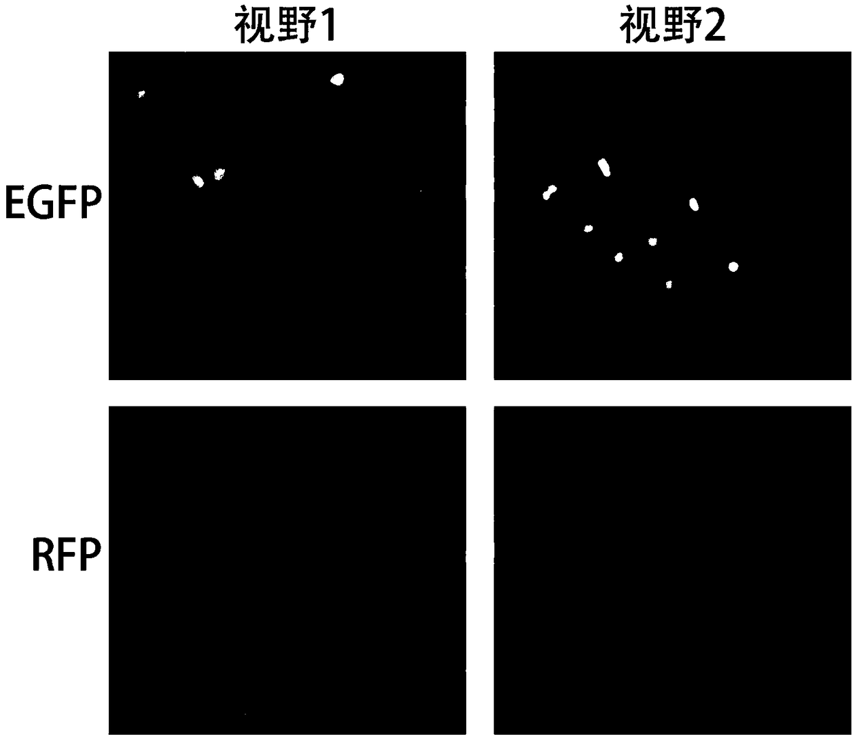
[0046] Passage 293FT cells to 6-well plates one day in advance, so that the cell density is 80% the next day. The next day for transfection: Take a new 1.5ml EP tube, add 100μL Opti-medium, then add 1μg of target plasmid, 0.5μg of packaging plasmid; take a new set of 1.5ml EP tube, add 5μl Liposome 2000 , gently flick the centrifuge tube to mix; after standing still for 5 minutes, add the plasmid mixture to the liposome mixture, flick the centrifuge tube gently to mix, and stand still for 30 minutes. The medium of the 293FT cells was replaced with 2 ml of fresh complete medium without antibiotics, and the liposome-plasmid complex was added. After 6 hours, the medium was changed again. After 48 hours of transfection, observe the fluorescence of 293FT cells, such asfigure 1 As indicated, 293FT cell culture supernatants were collected and centrifuged at 4°C for 5 min to remove cells. Filter the virus with a 0.45 μm filter, aliquot the viru...
Embodiment 3
[0048] Example 3 Obtaining Stable Nuclear Fluorescently Labeled Glioma Cells
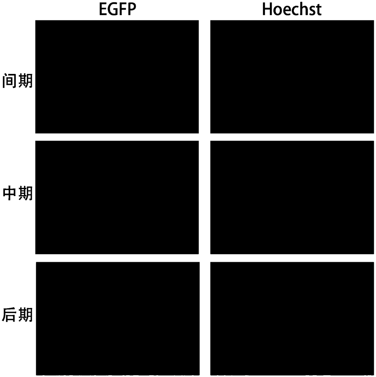
[0049] The glioma cells were passaged into 6-well plates one day in advance, so that the cell density was 30% the next day. Replace the medium of the cells in the 6-well plate. Add 500 μl of virus liquid into each 6-well plate, and add Polybrene at the same time, the final concentration is 8 μg / ml, and infect the cells overnight. Discard the medium containing the virus liquid and replace it with the normal complete medium. After obtaining a sufficient amount of cells one week later, the cells positive for EGFP and RFP were sorted by flow cytometry for expanded culture. At the same time, the cell slides were performed to observe the situation of the green fluorescently labeled nuclei, such as figure 2 shown. Obtain fluorescently labeled glioma cells.
PUM
 Abstract
Description
Claims
Application Information
 - R&D Engineer
- R&D Manager
- IP Professional
- Industry Leading Data Capabilities
- Powerful AI technology
- Patent DNA Extraction
Browse by: Latest US Patents, China's latest patents, Technical Efficacy Thesaurus, Application Domain, Technology Topic, Popular Technical Reports.
© 2024 PatSnap. All rights reserved.Legal|Privacy policy|Modern Slavery Act Transparency Statement|Sitemap|About US| Contact US: help@patsnap.com


