Oral squamous carcinoma cell strain as well as preparation method and applications thereof
A technology for oral squamous cell carcinoma and cancer cells, applied in the field of life medicine
- Summary
- Abstract
- Description
- Claims
- Application Information
AI Technical Summary
Problems solved by technology
Method used
Image
Examples
Embodiment 1
[0033] The mouse oral cancer oral cancer cell line is prepared according to the following method:
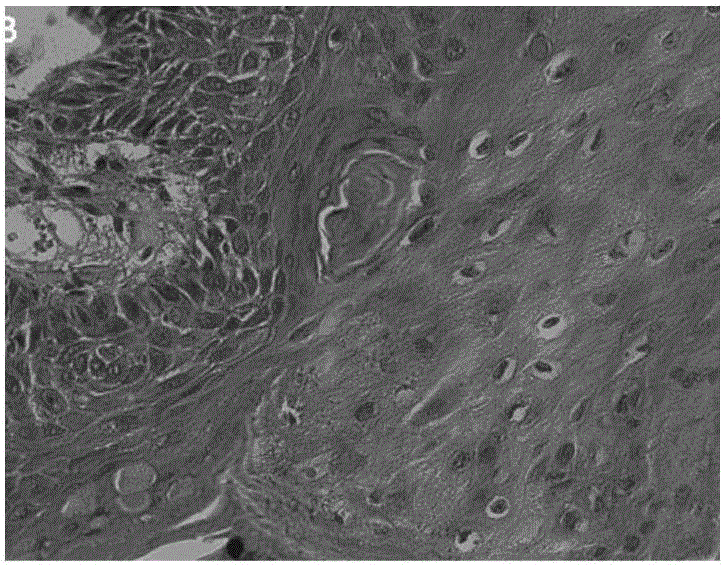
[0034] Step 1: Acquisition of mouse oral cancer tissue blocks: After drinking water to induce MAL gene knockout mice for 20 weeks with the conventional mouse oral cancer inducer 4NQO (80 μg / ml), the mucosa of the back of the tongue of a female mouse numbered 167# A cauliflower-shaped lesion was found, and the pathological diagnosis was oral squamous cell carcinoma, see figure 2 , use sterilized surgical instruments to quickly remove the tumor on the tip of the tongue, and use sterilized small scissors to quickly dissociate the tumor specimen, soak it in high-sugar DMEM solution containing 1% penicillin-streptomycin double antibody, and move it to a biological safety cabinet, containing double antibody The remaining tissue was obtained after repeatedly washing the tissue with sterile PBS buffer to remove impurities such as mucus and red blood cells;
[0035] Step 2: Tissue bloc...
Embodiment 2
[0040] Mice oral cancer cell line Mca-T-MAL of the present invention - / - detection and identification
[0041] 1. Morphological observation
[0042] Mainly observe the general morphology of cells, such as gross shape, nucleoplasmic ratio, chromatin and nucleolus size, number, etc., as well as the arrangement of cytoskeleton microfilaments and microtubules.
[0043] 1.1 Inverted optical microscope observation
[0044] The general morphology of cells, such as general shape, nuclear-to-cytoplasmic ratio, and adhesion characteristics, is mainly observed in the normal growth state.
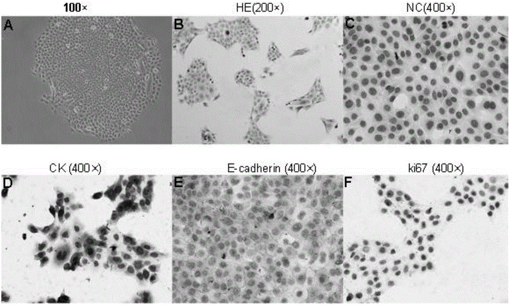
[0045] Observing the mouse oral cancer cell line Mca-T-MAL of the present invention under an inverted optical phase contrast microscope - / - , it can be seen that the cells are polygonal or elliptical, grow in groups, have large nuclei, abundant cytoplasm, tight connections between cells, and grow attached to the plate. HE staining shows that the nucleus is round, oval or kidney-shaped, the staining...
Embodiment 3
[0084] Mice oral cancer cell line Mca-T-MAL of the present invention - / - Establishment of in vivo animal models
[0085] 6-week-old BALB / C nude mice were selected for the experiment, half male and half male, and all animal experiments were carried out in accordance with the operating guidelines for animal experiments approved by Shanghai Jiaotong University School of Medicine.
[0086] 1. Subcutaneous tumor formation in nude mice
[0087] Step 1: The experimental cells are divided into two groups, which are respectively the cell line Mca-T-MAL of the present invention - / - and unselected primary mouse oral carcinoma cells;
[0088] Step 2: After cell digestion and counting, wash 2 times with serum-free and antibiotic-free DMEM, and dilute the cells to 1×10 with serum-free and double-antibody-free DMEM. 7 cells / ml of cell suspension, kept on ice;
[0089] Step 3: select two injection points for each nude mouse, inject 100 μl of cell suspension subcutaneously on the outer sid...
PUM
 Abstract
Description
Claims
Application Information
 - R&D Engineer
- R&D Manager
- IP Professional
- Industry Leading Data Capabilities
- Powerful AI technology
- Patent DNA Extraction
Browse by: Latest US Patents, China's latest patents, Technical Efficacy Thesaurus, Application Domain, Technology Topic, Popular Technical Reports.
© 2024 PatSnap. All rights reserved.Legal|Privacy policy|Modern Slavery Act Transparency Statement|Sitemap|About US| Contact US: help@patsnap.com


