SAA (serum amyloid A) immunochromatographic test strip and preparation method and test method thereof
An immunochromatographic detection and test strip technology, which is applied in the field of preparation of immunochromatographic detection test strips, can solve the problems of not asking the market, and achieve the effects of short detection time, high specificity, and high sensitive detection performance.
- Summary
- Abstract
- Description
- Claims
- Application Information
AI Technical Summary
Problems solved by technology
Method used
Image
Examples
Embodiment 1
[0048] Preparation of each component of the gold standard test paper of embodiment 1
[0049] 1 Preparation of colloidal gold solution
[0050] 0.01% HAuCl 4 Heat the solution to boiling, quickly add every 100mLHAuCl 4 The solution is added with an appropriate amount of reducing agent solution, the color changes from blue, then light blue, blue, then red after heating, and transparent orange-red after boiling for 7-10 minutes. Then filter with ultrafiltration or microporous membrane (0.45uM) to remove polymers and other possible impurities. The prepared colloidal gold should be pure, translucent, free of sediment and floating matter, and discard when oily matter and a large amount of black granular precipitate impurities appear on the liquid surface.
[0051] Wherein the reducing agent used can be trisodium citrate, tannic acid-trisodium citrate, white phosphorus, preferably use trisodium citrate, more preferably use 1% trisodium citrate. The glass containers used therein ...
Embodiment 2
[0075] Example 2 Preparation of colloidal gold-labeled anti-SAA antibody protein detection test paper 1-3
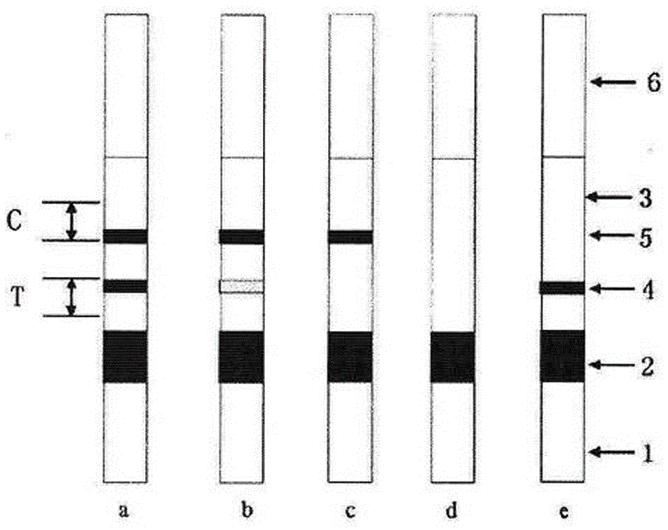
[0076] The sample pads prepared in Example 1 and dried, the binding pads, the analysis membrane, and the absorbent paper were cut into 1.7cm, 0.8cm, 2.5cm, and 1.5cm wide narrow strips with a cutting machine, according to figure 1 The way is overlapped into large slabs, and the large slabs are cut into single servings with a strip cutter. The width of each serving varies according to the jamming of the bottom plate. The preferred width of the present invention is 3mm. Assemble the test paper that has been cut for a single person into the prepared test paper card, so that the sample loading window corresponds to the sample pad of the test paper, and the result display window corresponds to the detection area and quality control area. 20~30%.
[0077] The components of gold standard test strips 1~3 are as follows:
[0078]
Embodiment 3
[0079] Embodiment 3 sample processing
[0080] Serum sample: Take 1~5mL of whole blood in a serum collection tube, let it stand for 30min~2h, centrifuge at 3000~5000g for 5~10min, and take the supernatant. According to the detection accuracy of the test strip, dilute the sample 0-100 times with the sample buffer solution, take 50-100uL dropwise into the sample hole of the test strip, and observe the result after standing for 5-20min.
[0081] Plasma sample: Take 1~5mL of whole blood, mix it in a sodium citrate or sodium heparin anticoagulant tube, centrifuge at 1000~3000g for 5~10min, and take the supernatant to get the plasma sample. According to the detection accuracy of the test strip, dilute the sample 0-100 times with the sample buffer solution, take 50-100uL dropwise into the sample hole of the test strip, and observe the result after standing for 5-20min.
[0082] Whole blood sample: Take about 50uL of fresh blood from the fingertip or earlobe, drop it into the sample ho...
PUM
| Property | Measurement | Unit |
|---|---|---|
| length | aaaaa | aaaaa |
| width | aaaaa | aaaaa |
Abstract
Description
Claims
Application Information
 - Generate Ideas
- Intellectual Property
- Life Sciences
- Materials
- Tech Scout
- Unparalleled Data Quality
- Higher Quality Content
- 60% Fewer Hallucinations
Browse by: Latest US Patents, China's latest patents, Technical Efficacy Thesaurus, Application Domain, Technology Topic, Popular Technical Reports.
© 2025 PatSnap. All rights reserved.Legal|Privacy policy|Modern Slavery Act Transparency Statement|Sitemap|About US| Contact US: help@patsnap.com


