Reagent box and detection method
The technology of a kit and a detection kit, which is applied in the field of breeding, can solve the problems of nucleic acid and primer reporting that have not yet been invented, and achieve the effects of rapid detection, high efficiency, and high detection sensitivity
- Summary
- Abstract
- Description
- Claims
- Application Information
AI Technical Summary
Problems solved by technology
Method used
Image
Examples
Embodiment 1
[0046] Example 1: Microsporidia PCR Rapid Detection Kit for Litopenaeus vannamei Intestinal Epithelial Cells
[0047] All the chemical reagents and primers of the microsporidia rapid detection kit for Litopenaeus vannamei intestinal epithelial cells described in this example were purchased from professional reagent companies. However, the sources of the above reagents and primers do not constitute any limitation to the present invention, and the present invention can prepare relevant reagents and synthesize relevant primers by itself.
[0048] The kit consists of the following parts (9 samples):
[0049] (1) 1.0mL 2×Reaction Mix Buffer, containing the following components:
[0050]
[0051] (2) Detection primers: upstream primer ECZ-F: 5'-TGTGGGAGAAATCTTAGTTTT-3', downstream primer ECZ-R: 5'-ATTGCGCTTGCTGCCCAGGAT-3', the concentration is 10 μM, the upstream and downstream primers are mixed, 400 μL.
[0052] (3) Taq enzyme 5U / μL.
[0053] (4) Sterilized ddH 2 O 1.0 mL. ...
Embodiment 2
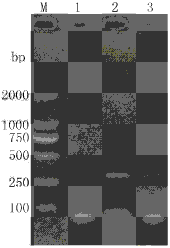
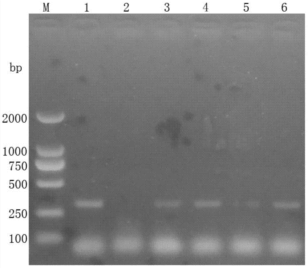
[0072] Example 2: PCR detection method for microsporidia in intestinal epithelial cells of Litopenaeus vannamei
[0073] Using the kit described in Example 1, proceed as follows:
[0074] (1) Take 50 mg of hepatopancreas and intestine samples of Litopenaeus vannamei to be tested, add 600 μL of sterilized double-distilled water, grind them thoroughly with a glass homogenizer, and place them in a -20°C refrigerator for three times of freezing and thawing. Centrifuge at low temperature at 6000rpm for 10 minutes, take the supernatant, add 200μL Tris-saturated phenol, shake and mix well, let stand for 5 minutes, then centrifuge at 12000rpm for 5 minutes, take the supernatant and add an equal amount of phenol: chloroform: isoamyl alcohol to extract Extract twice, take the supernatant and add 2 times the volume of isopropanol, mix well, let stand for 10 minutes, centrifuge at 12000rpm for 10 minutes, remove the supernatant, add 1mL pre-cooled 75% ethanol, let stand for 5 minutes, Ce...
Embodiment 3
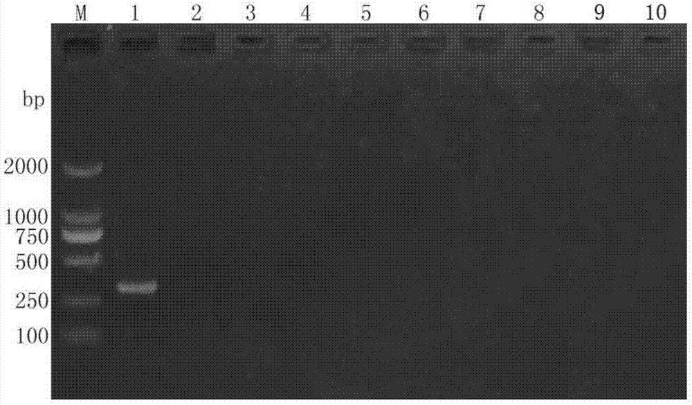
[0079] Example 3: Specificity experiment of PCR rapid detection kit for microsporidia in intestinal epithelial cells of Litopenaeus vannamei
[0080] Using the kit described in Example 1, proceed as follows:
[0081] (1) The extracted intestinal epithelial microsporidia, white spot syndrome virus, infectious subcutaneous and hematopoietic organ necrosis virus, prawn baculovirus, hepatopancreatic parvovirus, taura virus, yellow head virus, infectious muscle Necrosis virus, Vibrio parahaemolyticus, and Vibrio alginolyticus were used as templates for PCR detection.
[0082] (2) Take 10 μL of 2× reaction mixture buffer, 0.5 μL of upstream and downstream primers (ECZ-F, ECZ-R), 0.5 μL of Taq DNA polymerase, ddH 2 O 5.5 μL, template 3.0 μL. After mixing evenly, centrifuge for a few seconds and place on a PCR reaction instrument.
[0083] (3) Perform PCR amplification under the following conditions: 95°C for 5 min, 1 cycle; 94°C for 30 s, 53°C for 30 s, 72°C for 40 s, 30 cycles; 7...
PUM
 Abstract
Description
Claims
Application Information
 - R&D
- Intellectual Property
- Life Sciences
- Materials
- Tech Scout
- Unparalleled Data Quality
- Higher Quality Content
- 60% Fewer Hallucinations
Browse by: Latest US Patents, China's latest patents, Technical Efficacy Thesaurus, Application Domain, Technology Topic, Popular Technical Reports.
© 2025 PatSnap. All rights reserved.Legal|Privacy policy|Modern Slavery Act Transparency Statement|Sitemap|About US| Contact US: help@patsnap.com


