Electrogenerated chemiluminescence detection method of phospholipid and application of detection method
A technology of electrochemistry and luminescence detection, which is applied in the direction of chemiluminescence/bioluminescence, and analysis by making materials undergo chemical reactions, can solve the problems that cannot be applied to the detection of single-cell phospholipids, and achieve good specificity and detection sensitivity High, easy-to-operate effect
- Summary
- Abstract
- Description
- Claims
- Application Information
AI Technical Summary
Problems solved by technology
Method used
Image
Examples
Embodiment 1
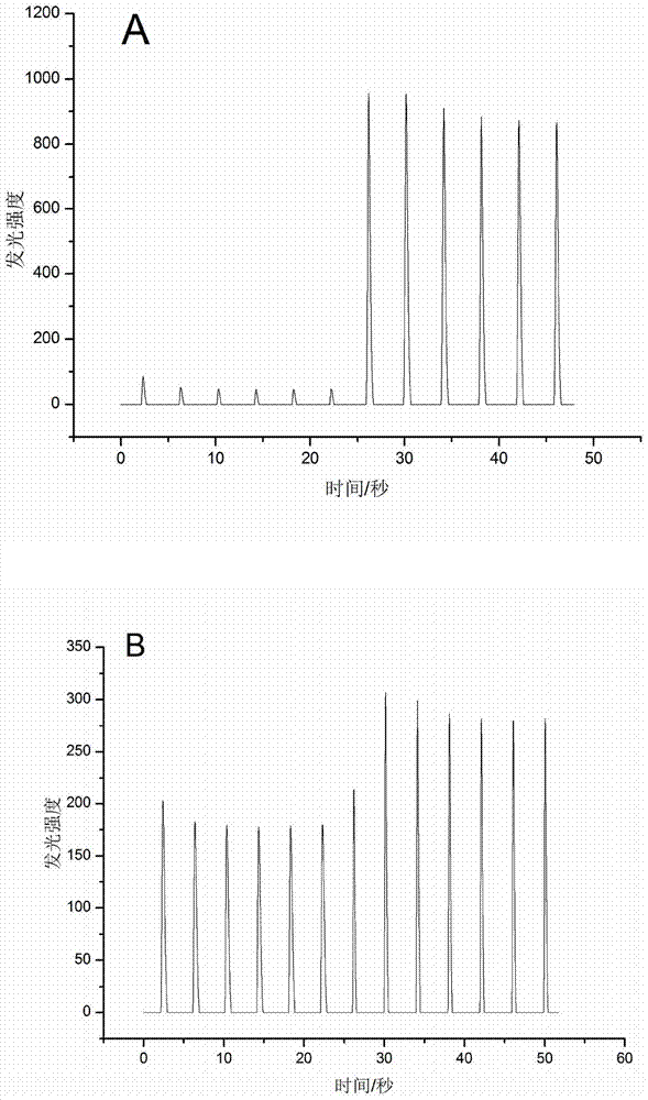
[0031] Example 1 Electrochemiluminescent detection of pure phospholipids.
[0032] First disperse the pure lecithin with chloroform, dry it with nitrogen in a fume hood, prepare a high-concentration lecithin solution, the solvent is pH=7.4PBS, and sonicate for 15 minutes to completely dissolve the sample. Using ITO as the working electrode, Ag / AgCl and Pt as the reference electrode and counter electrode respectively, an O-ring was attached to the surface of the ITO, and 200 μL of the solution to be tested (PBS solution containing 200 μM luminol and 1 mM lecithin) was added to it, The scanning speed is 1V / S, the voltage of -1.0V ~ 1.0V is applied to the ITO, the voltage of the photomultiplier tube is 500V, and the background luminous intensity I is recorded. 0 ;When recording the signal, try to ensure that there is no strong light shining on the instrument to avoid the error of the luminous intensity measured by the photomultiplier tube. After the baseline is stable, pause and ...
Embodiment 2
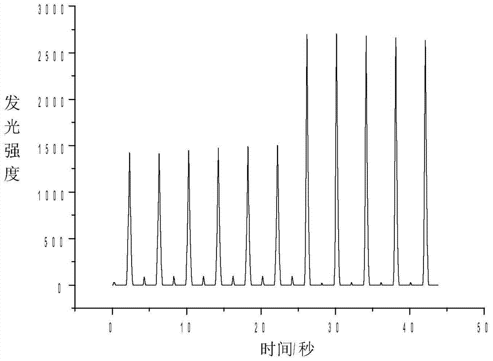
[0033] Example 2 Electrochemiluminescence detection of phospholipids on cell membrane surface.
[0034] (1) Cell membrane phospholipid activation: culture about 7000 Hela cells (with an area of 1 cm radius) on the surface of ITO, and after the cells grow on the wall overnight, use low ionic strength containing 0.5mM phosphate buffer (PBS, pH 7.4) and 310mM sucrose solution, incubated at 37°C for 1 hour to activate the phospholipids on the cell membrane surface, and then washed and soaked with 100mM PBS (pH 7.4).
[0035] (2) Detection: Take the ITO with cultured cells, suck off the low-ion solution, wash it with PBS for 3 times, add 190 μL of solution (PBS solution containing 200 μM luminol), ITO as the working electrode, Ag / Agcl and Pt As the reference electrode and the counter electrode respectively, the scanning speed is 1V / S during detection, the voltage is -1.0V~1.0V, the voltage of the photomultiplier tube is 600V, and the background luminous intensity is recorded. Aft...
Embodiment 3
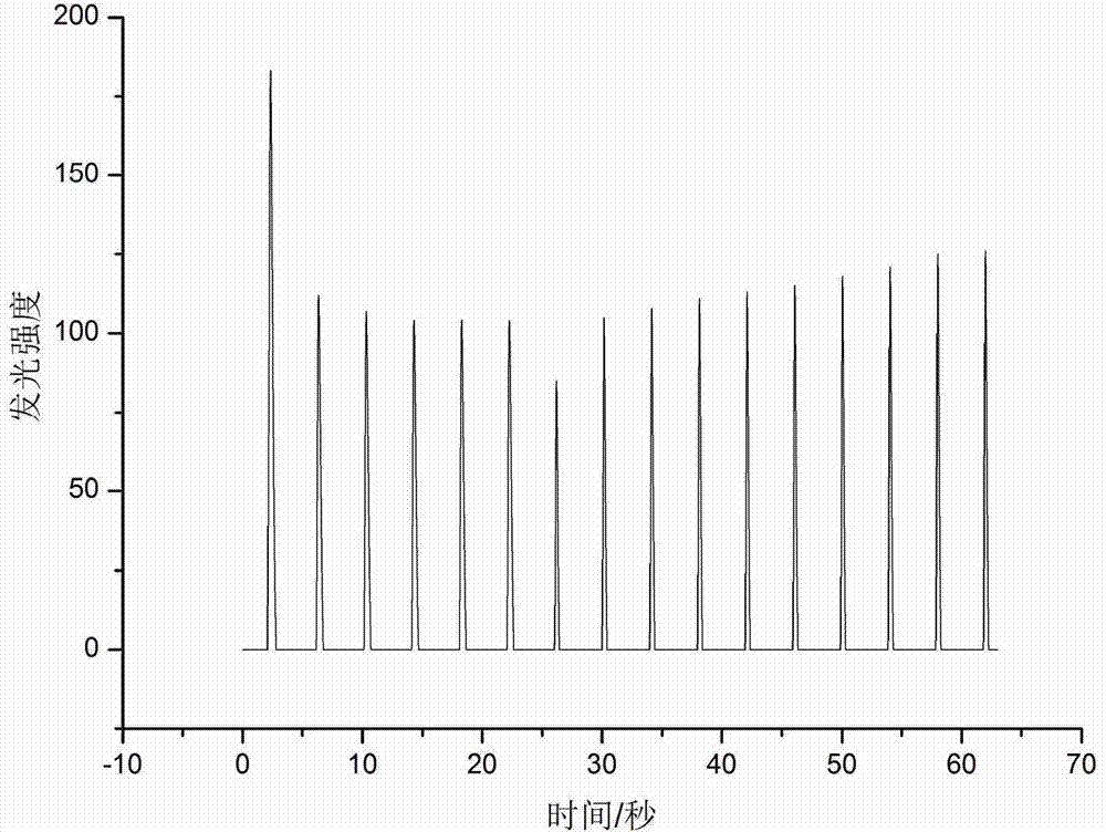
[0041] Example 3 Electrochemiluminescent detection of phospholipids on the surface of single cells.
[0042] For the detection of lipid molecules on the surface of single cells, first use pinhole technology to form a 30 μm single hole on the surface of ITO, treat the cultured Hela cells with trypsin, centrifuge, wash, and soak in a solution with low ionic strength, and finally put the single The cells are introduced into the single hole. Before the single cell is introduced, the ITO single hole must be vacuumed to make the single hole wet. Add the diluted cell solution from one side under the microscope, and then use absorbent paper from the other side to ensure that the cells are introduced into the ring-shaped single hole. Put the electrode into the incubator and culture it for about 1 hour to make the cells stick to the wall. After sticking to the wall, suck off the special high-glucose medium for the culture medium and add 190 μL of 200 μM luminol in PBS solution, so as to ...
PUM
 Abstract
Description
Claims
Application Information
 - R&D
- Intellectual Property
- Life Sciences
- Materials
- Tech Scout
- Unparalleled Data Quality
- Higher Quality Content
- 60% Fewer Hallucinations
Browse by: Latest US Patents, China's latest patents, Technical Efficacy Thesaurus, Application Domain, Technology Topic, Popular Technical Reports.
© 2025 PatSnap. All rights reserved.Legal|Privacy policy|Modern Slavery Act Transparency Statement|Sitemap|About US| Contact US: help@patsnap.com


