
A kind of elastic gel scaffold material for bone tissue engineering and its preparation method

A technology of bone tissue engineering and elastic gel, applied in the field of elastic gel scaffold materials, can solve the problems of loss of mechanical properties, easy dissolution, use of tissue engineering materials, etc., and achieve excellent biological functions and mechanical properties

Inactive Publication Date: 2015-10-28
NINGBO UNIVERSITY OF TECHNOLOGY
View PDF5 Cites 2 Cited by
  • Summary
  • Abstract
  • Description
  • Claims
  • Application Information

AI Technical Summary

Problems solved by technology

However, because divalent ions are easily exchanged with ions in the surrounding medium environment of the gel, the gel is easily dissolved in the body fluid environment and loses its mechanical properties.
In addition, due to its strong hydrophilicity, alginate has poor adhesion to proteins, and the lack of specific adsorption sites for cells limits the application of this gel.
[0007] At present, the existing double network gel system has good mechanical properties, but its cell adhesion is poor, it cannot grow on it, and it cannot be used as a tissue engineering material

Method used


Image

  • A kind of elastic gel scaffold material for bone tissue engineering and its preparation method
  • A kind of elastic gel scaffold material for bone tissue engineering and its preparation method
  • A kind of elastic gel scaffold material for bone tissue engineering and its preparation method

Examples


Embodiment 1

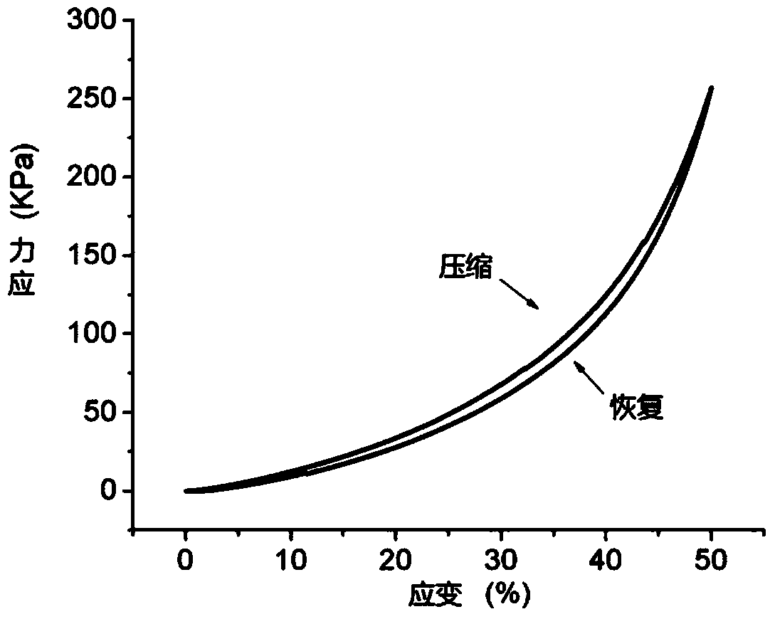
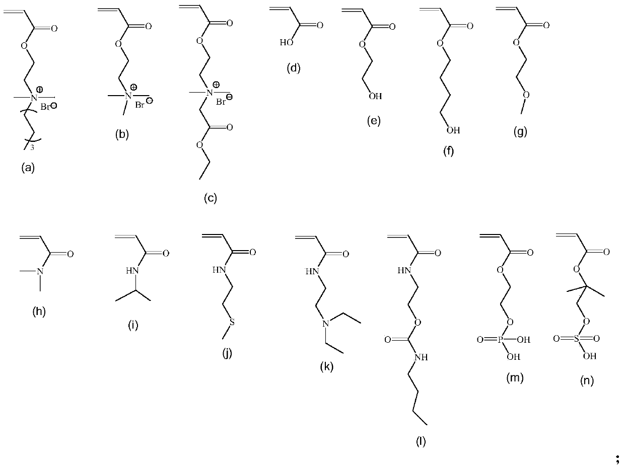
[0048] Step (1). Add 1.09 g of gel N1 reactive monomer (c), 0.54 g of gel N1 reactive monomer (h), and 0.024 g of N,N-methylenebisacrylamide cross-linked monomer into a 15 ml plastic tube. The joint agent and tetramethylethylenediamine accelerator were dissolved in 6.5 ml of deionized water, and nitrogen gas was introduced for 30 minutes to remove the oxygen in the system, and then 300 microliters of ammonium persulfate solution (1% in mass) was added with a syringe, Shake well and react at 25°C for 12 hours to obtain gel N1.

[0049] Step (2). Take the gel N1 out of the plastic tube and cut it into 1-3 cm columnar gel blocks with a knife. Prepare a 200 mL gel N2 reaction solution of acrylamide (28.4 g), α-ketoglutarate (0.58 g), and N,N-methylenebisacrylamide (0.062 g). Put the gel block of gel N1 into the gel N2 reaction solution, seal it with aluminum foil, and shake slightly for 48 hours. Then, take out the gel block that has been fully soaked, and put it under ultraviol...

Embodiment 2

[0052] Step (1). Add 1.09 g of gel N1 reactive monomer (c), 0.54 g of gel N1 reactive monomer (h), and 0.024 g of N,N-methylenebisacrylamide cross-linked monomer (c) into a 15 ml plastic tube. The joint agent and tetramethylethylenediamine accelerator were dissolved in 6.5 ml of deionized water, and nitrogen gas was introduced for 30 minutes to remove the oxygen in the system, and then 300 microliters of ammonium persulfate solution (1% in mass) was added with a syringe, Shake well and react at 25°C for 12 hours to obtain gel N1.

[0053] Step (2). Take the gel N1 out of the plastic tube and cut it into 1-3 cm columnar gel blocks with a knife. Prepare a 200 mL gel N2 reaction solution of acrylamide (56.8 g), α-ketoglutarate (1.16 g), and N,N-methylenebisacrylamide (0.124 g). Put the gel block of gel N1 into the gel N2 reaction solution, seal it with aluminum foil, and shake slightly for 48 hours. Then, take out the gel block that has been fully soaked, and put it under the u...

Embodiment 3

[0055] The double network gel obtained in Example 2 was cut into small pieces of 5mm*5mm*2mm, washed three times with deionized water, frozen at -20°C, and dried in vacuum, then soaked in 70% alcohol solution After 2 hours, wash with PBS for 3 times in a sterile environment, and then soak overnight in PBS.

[0056] Adipose-derived mesenchymal stem cells were planted on the double network gel, added with DMEM growth medium containing 10% fetal bovine serum, cultured at 37°C, 5% carbon dioxide environment for 3 days, and stained with DAPI dye. A large number of adipose stem cells adhered and grew on the surface of the gel, which indicated that the gel had excellent biocompatibility. When the fullness increased to about 80%, the osteoinductive medium (StemPro, GIBCO) was used to culture at 37°C and 5% carbon dioxide for 21 days. The double network gel scaffold was stained with alizarin, and the calcified morphology of the gel surface could be observed under a microscope. After ...


PUM

PropertyMeasurementUnit
elastic modulusaaaaaaaaaa

Abstract

The invention discloses an elastic gel support material for bone tissue engineering and a preparation method of the material. The material consists of a gel N1 and a gel N2, wherein the molar content of the gel N1 is 5-50% and the molar content of the gel N2 is 50-95%. The preparation method comprises the following steps: fully dissolving a gel N1 reaction monomer, a gel N1 crosslinking agent and a gel N1 accelerant in deionized water to obtain a gel N1 synthesized solution; introducing N2 to remove oxygen in the solution, and adding a free radical initiator to initiate polymerization of the free radical to obtain the gel N1; fully dissolving a gel N2 reaction monomer, a gel N2 crosslinking agent and a photoinitiator in deionized water to obtain a gel N2 synthesized solution; and adding the gel N1 into the gel N2 synthesized solution to be fully immersed, and then irradiating and photo-initiating polymerization of the free radical under an ultraviolet lamp to obtain a double-network gel. The component of gel in the material is cytoactive, can adapt in vivo dynamic environment and can be applied to bone tissue repair materials.

Description

technical field [0001] The invention relates to a hydrogel tissue engineering material, in particular to an elastic gel support material for bone tissue engineering, which can be used as a carrier for cartilage repair and drug controlled release system. Background technique [0002] The regeneration ability of cartilage tissue is poor, and the repair of cartilage defect is a difficult problem in clinical medicine. At present, autograft or allograft is mainly used for repair. Autologous transplantation has the disadvantages of less donor sources, which can easily lead to deformity and infection of the donor site after surgery, while allogeneic cartilage transplantation also has the problems of less supply sources and immune diseases, and it is difficult for both of them to achieve satisfactory results. Using tissue engineering methods to prepare biocompatible cell scaffolds and perform cell transplantation to repair tissues is a new way to solve this problem. [0003] Gels ...

Claims


Application Information

Patent Timeline
Patent Type & Authority Patents(China)
IPC IPC(8): A61K31/00A61L27/18A61L27/52A61L27/38C08F220/34C08F222/22C08F220/06C08F220/28
Inventor 蒋志强杨建邵双喜顾群
Owner NINGBO UNIVERSITY OF TECHNOLOGY
Learn More