Efficient Gd-loaded liposome preparation and preparation method thereof
A technology of gadolinium-loaded lipids and liposomes, which is applied in preparations for in vivo tests, pharmaceutical formulations, nuclear magnetic resonance/magnetic resonance imaging contrast agents, etc., to achieve small particle size, good stability, and good sustained-release effect Effect
- Summary
- Abstract
- Description
- Claims
- Application Information
AI Technical Summary
Problems solved by technology
Method used
Image
Examples
Embodiment 1
[0029] Weigh 10g of soybean lecithin, 2.5g of cholesterol, and 0.2g of vitamin E, and dissolve them in 300ml of chloroform, remove the solvent by rotary evaporation under constant temperature and reduced pressure at 45°C to form a lipid film; add 1000ml of phosphate buffer (pH7.4) for hydration Elute the film for 3.5 hours to obtain blank liposomes; carry out high-pressure homogenization of the blank liposomes, homogenize 15 times at 50 MPa, filter and sterilize with a 0.22 micron filter membrane, and vacuum concentrate to 20 ml at room temperature to obtain Phospholipid gel. Take 1ml of phospholipid gel and mix it with 1ml of 0.8mmol / ml Gd-DTPA, and incubate at 45°C for 5 hours. Packing and sealing.
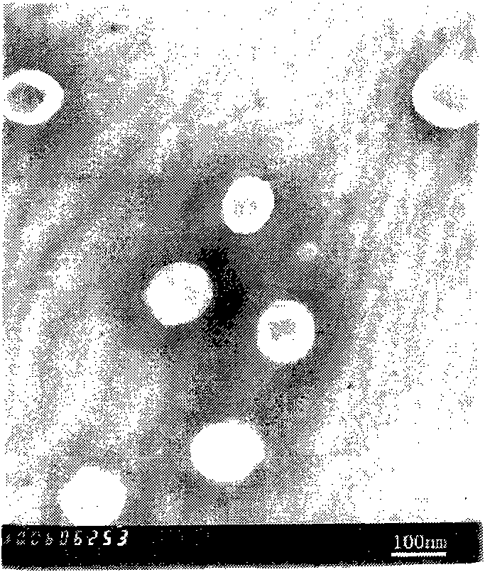
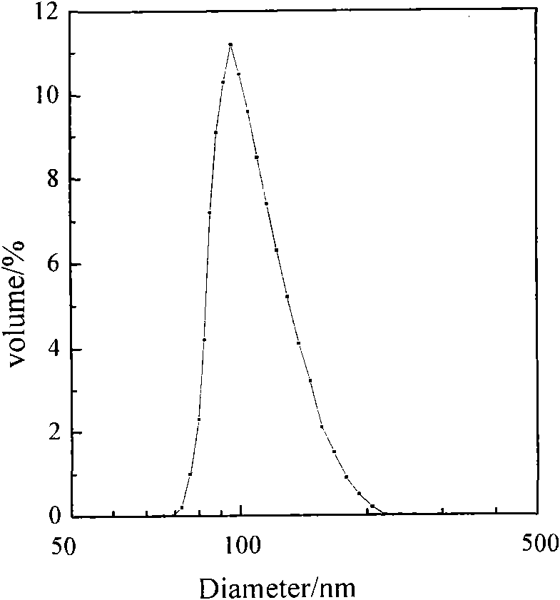
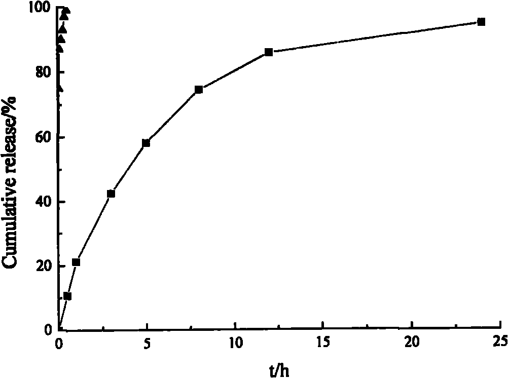
[0030] The volume average particle diameter measured by a dynamic light scattering laser particle size analyzer is 107.0nm, and its transmission electron microscope pictures and particle size distribution diagrams are shown in figure 1 and figure 2 , its in vitro sustained r...
Embodiment 2
[0034] Weigh 24g of egg yolk phospholipids, 4g of cholesterol, and 0.6g of vitamin E, and dissolve them in 300ml of chloroform and methanol (volume ratio 1:1), remove the solvent by rotary evaporation at a constant temperature and reduced pressure at 35°C to form a lipid film; add 1500ml of normal saline for hydration Elute the film for 3 hours to obtain blank liposomes; homogenize the blank liposomes under high pressure, 10 times at 80 MPa, filter and sterilize with a 0.22 micron filter membrane, and vacuum concentrate to 20 ml at room temperature to obtain phospholipids gel. Take 1ml of phospholipid gel and mix with 1ml of 0.6mmol / ml Gd-DTPA-BMA, and incubate at 45°C for 5 hours. Packing and sealing.
Embodiment 3
[0036] Weigh 15g of hydrogenated soybean lecithin, 2g of cholesterol, and 0.4g of vitamin E, and dissolve them in 300ml of absolute ethanol, remove the solvent by rotary evaporation at a constant temperature and reduced pressure at 40°C to form a lipid film; add 1000ml of phosphate buffer (pH6.8) for Hydrate and elute the film for 3 hours to prepare blank liposomes; carry out high-pressure homogenization of the blank liposomes, homogenize 6 times at 90 MPa, filter and sterilize with a 0.22 micron filter membrane, and vacuum concentrate to 20 ml at room temperature to obtain Phospholipid gel. Take 1ml of phospholipid gel and mix with 1ml of 0.4mmol / ml Gd-DTPA, and incubate at 45°C for 5 hours. Packing and sealing.
PUM
| Property | Measurement | Unit |
|---|---|---|
| Volume average particle size | aaaaa | aaaaa |
Abstract
Description
Claims
Application Information
 - R&D
- Intellectual Property
- Life Sciences
- Materials
- Tech Scout
- Unparalleled Data Quality
- Higher Quality Content
- 60% Fewer Hallucinations
Browse by: Latest US Patents, China's latest patents, Technical Efficacy Thesaurus, Application Domain, Technology Topic, Popular Technical Reports.
© 2025 PatSnap. All rights reserved.Legal|Privacy policy|Modern Slavery Act Transparency Statement|Sitemap|About US| Contact US: help@patsnap.com


