
Implantable device for penetrating and delivering agents to cardiac tissue

a technology of implantable devices and cardiac tissue, which is applied in the field of endocardial mapping, can solve the problems of premature stimuli to some regions of the heart, more complicated spatial and temporal disruption of the electrical synchronization of the heart cells, and completely disrupting the action of the heart overall, so as to effectively eliminate arrhythmogenic sites, reduce the effect of arrhythmogenic effects, and reduce the amount of damage upon implantation

Inactive Publication Date: 2001-12-11
BIOCARDIA
View PDF21 Cites 143 Cited by
  • Summary
  • Abstract
  • Description
  • Claims
  • Application Information

AI Technical Summary

Benefits of technology

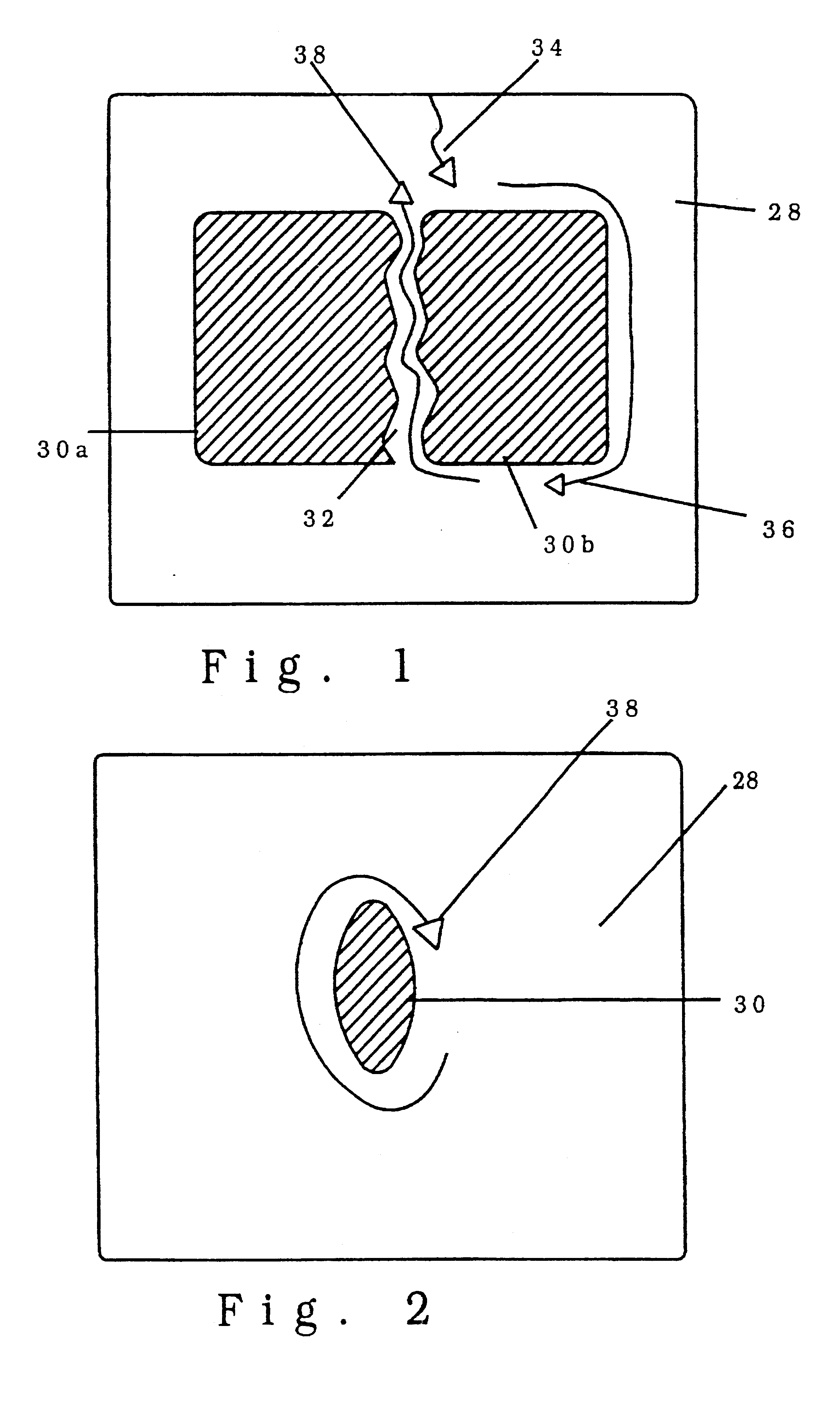
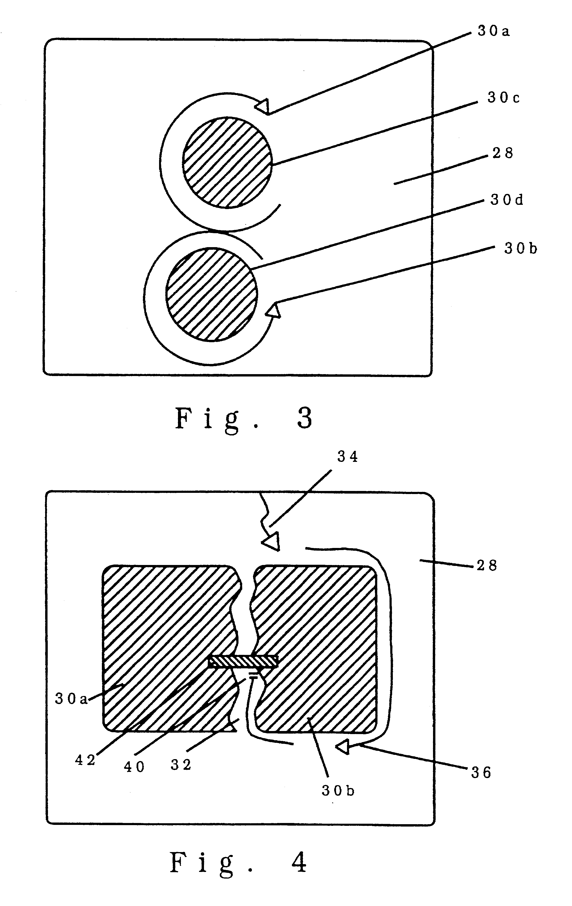
In general it is an object of the present invention to provide an implantable biocompatible device, and means for implantation of the device in the region of an arrhythmogenic site to effectively eliminate arrhythmogenic sites from the myocardium.
Another object of the invention is to provide a system and method of the above character that introduces little damage upon implantation such that it is essentially reversible. A reversible procedure allows an electrophysiologist the option to effectively interrupt, isolate, or otherwise effectively electrically remove or reduce the arrhythmogenic effects of a region of the myocardium that is believed to be the arrhythmogenic site, to perform evaluative tests if desired, and to modify the procedure only if desired based upon the results of the test or tests performed.
Another object of the invention is to provide a system and method of the above character in which the electrical disruptions introduced by the device into the myocardium are accurate and repeatable such that the effects on the arrhythmogenic sites in the myocardium may be introduced accurately and repeatably.
Another object of the invention is to provide a system and method of the above character in which the effects introduced into the myocardium may be altered by selecting different conductive and non conductive materials.
Another object of the invention is to provide a system and method of the above character in which the electrical disruptions introduced by the device into the myocardium are accurate and repeatable such that the effects on the arrhythmogenic sites in the myocardium may be introduced accuracy and repeatably.
Another object of the invention is to provide a system and method of the above character in which the effects introduced into the myocardium may eliminate arrhythmias at any depth within the heart tissue.

Problems solved by technology

In turn, such physiological disturbances introduce more complicated spatial and temporal disruption of the electrical synchronization of the heart cells necessary for proper heart function.
These inappropriate conduction pathways result in premature stimuli to some region of the heart by bypassing the normal conduction pathways.
Such premature depolarization of surrounding heart cells on a small scale is often sufficient to completely disrupt the action of the heart overall.
For example, in a myocardial infarction, or heart attack, cells die due to lack of nutrients because the blood vessel that provides the nutrients is obstructed.
As the site of infarction heals, the dead myocardium is replaced by fibrous tissue and the residual viable myocardial cells become embedded in scar leading to non-uniform activation and slow conduction.
Necrotic regions that act as arrhythmogenic sites may depend upon other arrhythmogenic sites to introduce an arrhythmia, just as the presence of other arrhythmogenic sites may complicate an arrhythmia.
This technique is hazardous for the patient in that it requires open heart surgery.
The procedure is complex in that it required a number of precisely located delicately introduced incisions in the heart wall.
The procedure is innovative in that it may result in a cure, but it is expensive and risky for the patient due to its complexity.
In these procedures, the arrhythmogenic region is isolated or the inappropriate pathway is disrupted by destroying the cells in the regions of interest.
A typical procedure involves the destruction of much more tissue than that required to terminate the arrhythmia, and much of this is unnecessary damage.
Since the dead tissue cannot be restored after each evaluation, the procedure is irreversible.
This temporary treatment is too short to allow for any but the most cursory evaluation of results.
In addition, if the temporary cooling is deemed successful, the procedure must be repeated allowing the introduction of procedural errors.
In addition, existing ablation techniques which involve complicated energy transfer mechanisms require approximations on the amount of myocardial tissue necrosis resulting from the destructive energy transfer mechanism used.
Repeatability and reliability of a procedure that varies inherently is extremely difficult.
Such mechanisms are also often limited in that they cannot eliminate tissue at a specified depth within the myocardium, but rather must typically begin destruction at the surface of the endocardium and move inwards until the desired depth is achieved.
One of the primary limitations of ablation techniques is the inability of the thermal action of RF energy to penetrate to arrhythmogenic sites deep in the heart wall.
Existing ablation techniques have a further drawback in that they may generate bubbles or introduce thrombosis formation in the heart.
However, bubbles or thrombosis could very easily be fatal or introduce brain damage if introduced in the left heart chambers.
They do not provide for local delivery of the drug to the heart, but only for controlled release into the gastro intestinal tract.
.]This work shows the viability of controlled release therapy delivered locally for the treatment of arrhythmias, but no means of introducing the agents directly to the arrhythmogenic site has been developed.

Method used


Image

  • Implantable device for penetrating and delivering agents to cardiac tissue
  • Implantable device for penetrating and delivering agents to cardiac tissue
  • Implantable device for penetrating and delivering agents to cardiac tissue

Examples


Embodiment Construction

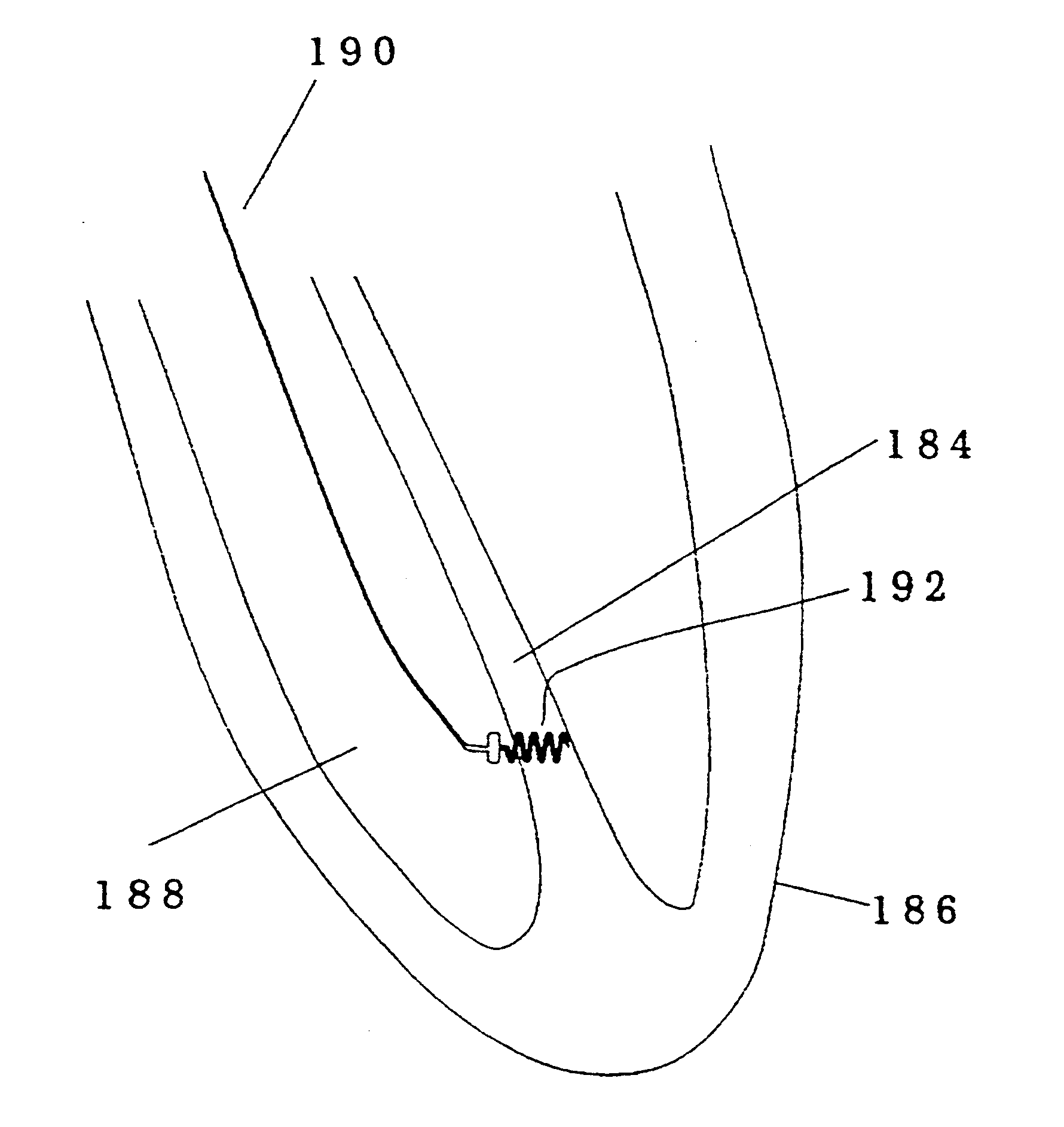
FIG. 7 shows a perspective view of a helix embodiment of this implantable device. In this preferred embodiment, the entire structure is made of a biocompatible Platinum Iridium alloy that can be formed using investment casting, machining or other similar techniques. Helix embodiment shown has a sharp tip 44 to allow for ease in advancing the helical structure into the heart wall. A number of loops of a helix 46 have the same diameter 48 and spacing 50 to prevent excessive damage to the myocardium upon insertion. By having the same diameter 48 at each cross section of the helix 46 and spacing 50 between the loops of the helix 46 the path through the myocardium followed by each loop of the helix 46 will be the same. The spacing 50 can vary from very tight spacing in which distance 50 between the two loops of helix 46 are approximately two times the size of diameter 52 of structure 53 that defines helix 46 to loose spacing in which distance 50 between the two loops of helix 46 are appr...


PUM


Abstract

An implantable devices for the effective elimination of an arrhythmogenic site from the myocardium is presented. By inserting small biocompatible conductors and / or insulators into the heart tissue at the arrhythmogenic site, it is possible to effectively eliminate a portion of the tissue from the electric field and current paths within the heart. The device would act as an alternative to the standard techniques for the removal of tissue from the effective contribution to the hearts electrical action which require the destruction of tissue via energy transfer (RF, microwave, cryogenic, etc.). This device is a significant improvement in the state of the art in that it does not require tissue necrosis.In one preferred embodiment the device is a non conductive helix that is permanently implanted into the heart wall around the arrhythmogenic site. In variations on the embodiment, the structure is wholly or partially conductive, the structure is used as an implantable substrate for anti arrhythmic, inflammatory, or angiogenic pharmacological agents, and the structure is deliverable by a catheter with a disengaging stylet. In other preferred embodiments that may incorporate the same variations, the device is a straight or curved stake, or a group of such stakes that are inserted simultaneously.

Description

BACKGROUND--FIELD OF INVENTIONThis invention relates to the field of endocardial mapping, and more particularly to the new field of devices for non-destructive elimination of arrhythmogenic sites and inappropriate conduction pathways, catheter methods for implantation of such devices and the use of such devices as substrates for local controlled drug release therapy.BACKGROUND--PRIOR ARTCardiac arrhythmias are abnormal rhythmic contractions of the myocardial muscle, often introduced by electrical or irregularities in the heart tissue. A region of the heart that results in an arrhythmia is here defined as an arrhythmogenic site in that it introduces the arrhythmia. If a number of regions acting in unison introduce an arrhythmia, they are each considered arrhythmogenic sites. Types of arrhythmogenic sites include, but are not limited to: accessory atrioventricular pathways, ectopic foci, and reentrant circuits.The anatomical causes of heart arrhythmias are numerous and not entirely un...

Claims


Application Information

Patent Timeline
Patent Type & Authority Patents(United States)
IPC IPC(8): A61F2/00A61N1/05
CPCA61F2/00A61N1/056A61N1/0573
Inventor ALTMAN, PETER A.
Owner BIOCARDIA
Learn More