
Method and device for mass spectrometric analysis of biomolecules using charge transfer dissociation (CTD)

a mass spectrometric and biomolecule technology, applied in the direction of electrical equipment, electric discharge tubes, particle separator tubes, etc., can solve the problems of unreasonably long analysis time for peptide sequencing and identification, particular limitation of hydrogen/deuterium scrambling, and obvious problems in mobilization

Active Publication Date: 2018-06-12
WEST VIRGINIA UNIVERSITY
View PDF3 Cites 0 Cited by
  • Summary
  • Abstract
  • Description
  • Claims
  • Application Information

AI Technical Summary

Benefits of technology

The patent text describes a new method called CTD (Cathode Ray Tube Discharge) for analyzing oligosaccharides using a modified ion trap mass spectrometer. CTD is compared to other methods like XUV-PD and CPD for analyzing the same sugar molecules, and it is found that CTD is more practical and affordable than other high-energy tandem MS methods. The method uses high-energy collisions to break down the sugar molecules and produce various fragments. The resulting fragments can be used to determine the complex structure of modified glycans. Interestingly, the method also has a similar mechanism to EID (Electron Ion Dissociation) and MAD (Matrix Assisted Laser Desorption Ionization) methods, which indicates that it may involve the same process of radical cleavages. The results from this study have potential applications in the field of glycan analysis.

Problems solved by technology

Although per-residue deuterium incorporation can be measured from the comparison of two peptides that differ by one residue in length, highly digested samples create complicated datasets that result in unreasonably long analysis times for peptide sequencing and identification.22,23
Although CID combined with HDX has shown some success,30,32 a particular limitation is hydrogen / deuterium (HD) scrambling.
A problem with CID is that it is accompanied by the mobilization of protons.33 These mobile protons, found on both acidic and basic residues, can migrate throughout the molecule and participate in the fragmentation process.33 Because proton mobilization occurs before dissociation in CID, the final location of the proton on the product ion is typically different than the initial location on the unactivated precursor ion.
Mobilization is obviously problematic when using HDX-MS / MS to target structural areas because redistribution occurs equally for mobile deuteriums as it does for mobile hydrogens.
In such cases, per-residue measurements provide ambiguous or erroneous structural information.
Given that HDX-MS platforms produce often produce peptides in low charge states (i.e. 1+ or 2+) using electrospray ionization, per-residue measurements can be difficult to obtain.

Method used


Image

  • Method and device for mass spectrometric analysis of biomolecules using charge transfer dissociation (CTD)
  • Method and device for mass spectrometric analysis of biomolecules using charge transfer dissociation (CTD)
  • Method and device for mass spectrometric analysis of biomolecules using charge transfer dissociation (CTD)

Examples


example 1

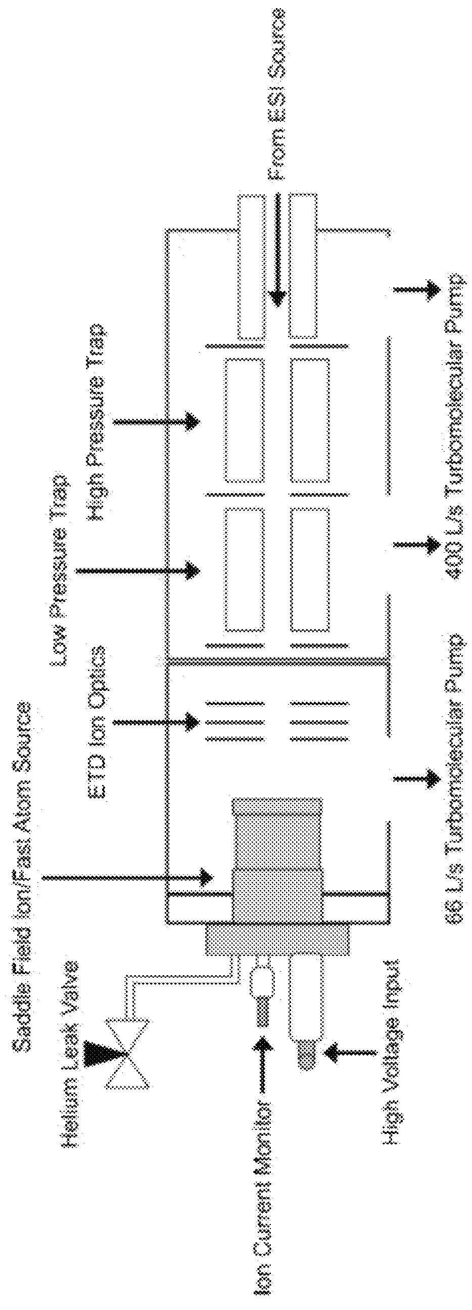
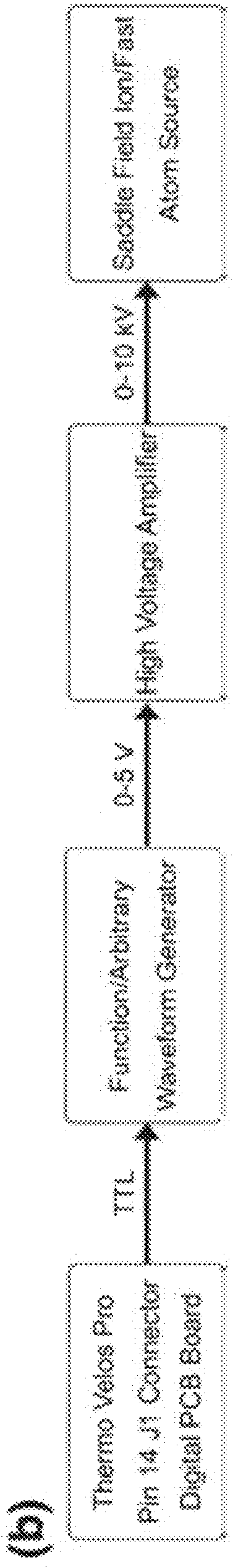
[0094]The Experimental setup is shown schematically in FIG. 1. A custom fabricated rear cover was mounted to the saddle field source along the axis of the ETD source ion optics. Briefly, a saddle field fast ion / fast atom source with an ion gun cathode in place, was interfaced to the ETD chamber of an LQT Velos Pro (Thermo Electron Corporation, San Jose, Calif., USA) mass spectrometer using a home built vacuum chamber cover. A variable leak valve was used to control the flow of helium through the addle field source. A 6 kV waveform from a high voltage amplifier was applied to the reagent ion source during the scan function normally reserved for CID, which was similar to previous MAD-MS experiments. FIG. 2 shows the routing of signals from the mass spectrometer to the saddle field ion / fast atom source (VSW / Atomtech, Inc. Macclesfield, UK). The trigger source was taken from pin 14 on the J1 connector of the Digital PCB board. This TTL signal was used to trigger an arbitrary waveform ge...

example 2

[0106]CTD mass spectrometry analysis of carbohydrates was also performed. Briefly, CTD mass spectrometric analysis was performed on oligosaccharides (carbohydrates) using mass spectrometric methods described in Example 1. The results are demonstrated in FIGS. 8-12. The results demonstrate that CTD can be used to sequence modified oligosaccharides and identify the location of the modifications.

Example 3. CTD Mass Spectrometry of Peptide Cations: Charge State Dependence and Side-Chain Losses

[0107]Introduction. In recent years, mass spectrometry (MS) has become an indispensable tool for the study of biological molecules such as lipids [1], oligosaccharides [2], peptides [3, 4], proteins [5], and DNA [6]. With the development of soft ionization methods such as fast atom bombardment (FAB), matrix-assisted laser desorption / ionization (MALDI) and electrospray ionization (ESI), single-stage MS plays an important role in the molecular weight determinations of an intact molecule of interest [...

example 3

REFERENCES FOR EXAMPLE 3

[0150]1. Lee, H.; An, H. J.; Lerno, L. A.; German, J. B.; Lebrilla, C. B.: Rapid profiling of bovine and human milk gangliosides by matrix-assisted laser desorption / ionization fourier transform ion cyclotron resonance mass spectrometry. International Journal of Mass Spectrometry 305, 138-150 (2011)[0151]2. Ko, B. J.; Brodbelt, J. S.: 193 nm ultraviolet photodissociation of deprotonated sialylated oligosaccharides. Analytical Chemistry 83, 8192-8200 (2011)[0152]3. Lopez-Clavijo, A. F.; Duque-Daza, C. A.; Creese, A. J.; Cooper, H. J.: Electron capture dissociation mass spectrometry of phosphopeptides: Arginine and phosphoserine. International Journal of Mass Spectrometry 390, 63-70 (2015)[0153]4. Voinov, V. G.; Hoffman, P. D.; Bennett, S. E.; Beckman, J. S.; Barofsky, D. F.: Electron capture dissociation of sodium-adducted peptides on a modified quadrupole / time-of-flight mass spectrometer. Journal of the American Society for Mass Spectrometry 26, 2096-2104 (201...


PUM


Abstract

Provided herein are devices, systems, and methods of CTD mass spectrometry analysis of biomolecules.

Description

[0001]This application claims the benefit of and priority to U.S. Provisional Patent Application No. 62 / 220,305, filed on Sep. 18, 2015, entitled “METHOD AND DEVICE FOR MASS SPECTROMETRIC ANALYSIS OF BIOMOLECULES USING CHARGE TRANSFER DISSOCIATION (CTD),” the contents of which is incorporated by reference herein in its entirety.STATEMENT REGARDING FEDERALLY SPONSORED RESEARCH OR DEVELOPMENT[0002]This invention was made with government support under grant number 1R01GM114494-01 awarded by the National Institutes of Health. The government has certain rights in the invention.BRIEF DESCRIPTION OF THE DRAWINGS[0003]Further aspects of the present disclosure will be readily appreciated upon review of the detailed description of its various embodiments, described below, when taken in conjunction with the accompanying drawings.[0004]FIG. 1 shows one embodiment of a mass spectrometer configured to separate sample ions via charge transfer dissociation using helium cations.[0005]FIG. 2 shows a ...

Claims


Application Information

Patent Timeline
IPC IPC(8): H01J49/26H01J49/00
CPCH01J49/0072
Inventor JACKSON, GLEN P.HOFFMANN, WILLIAM D.
Owner WEST VIRGINIA UNIVERSITY
Learn More