Therapeutic applications of calcium electroporation to effectively induce tumor necrosis
a technology of electroporation and therapy, applied in the field of calcium overload, can solve the problems that its use in cancer treatment has not been fully investigated, and achieve the effects of reducing the amount of surgery, high efficiency, and increasing the intracellular calcium level through-ou
- Summary
- Abstract
- Description
- Claims
- Application Information
AI Technical Summary
Benefits of technology
Problems solved by technology
Method used
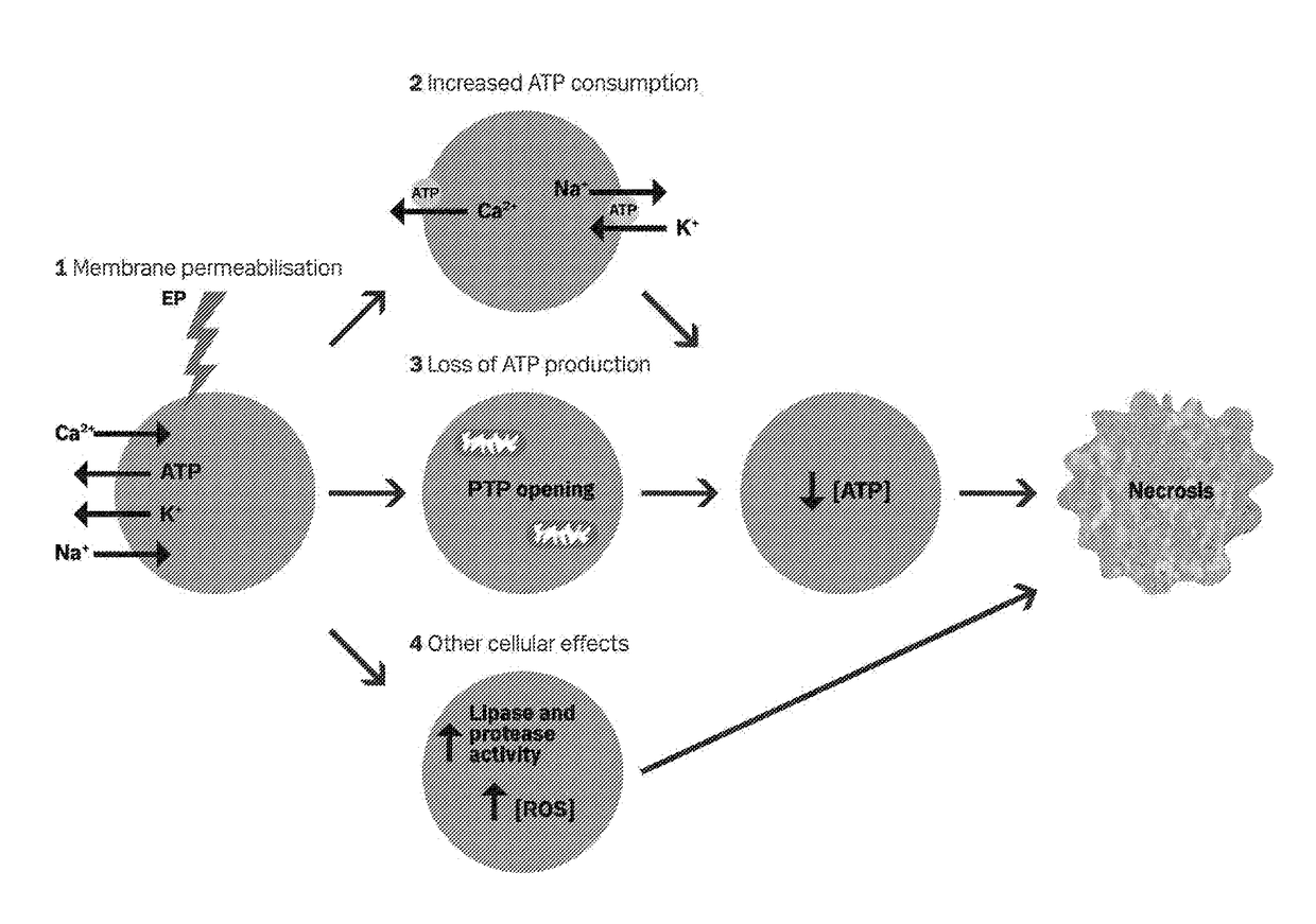
Image

Examples
example 1
Materials and Methods
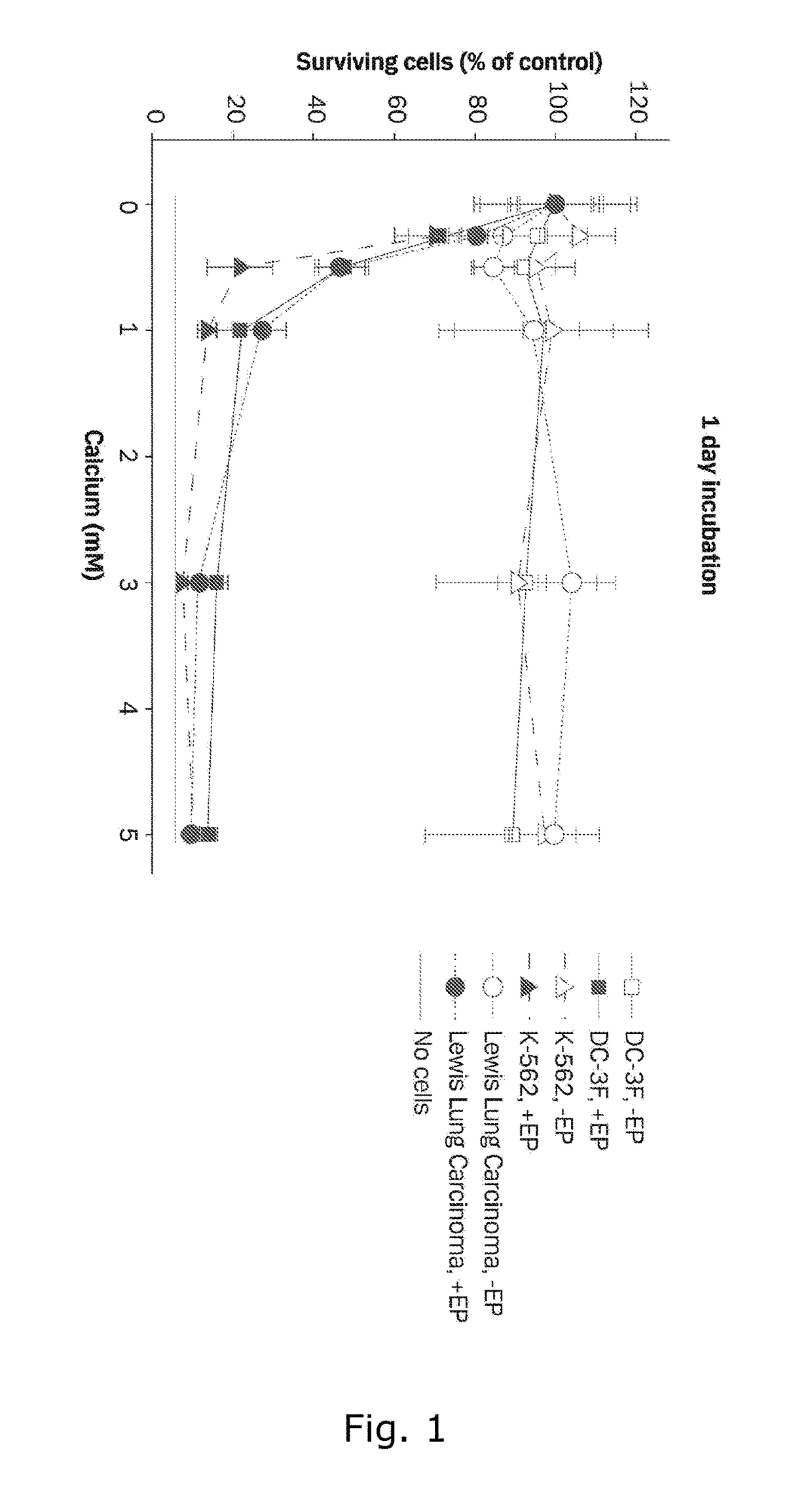
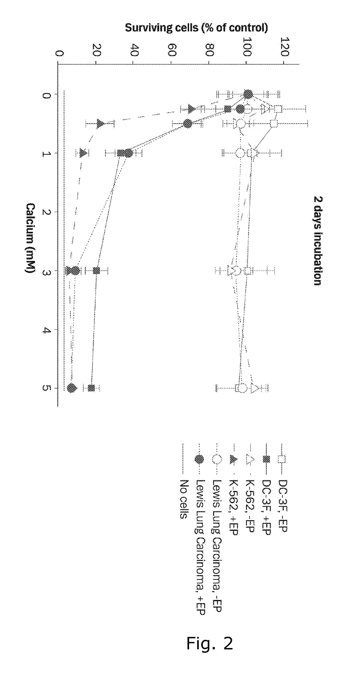
[0175]Three cell lines are used for in vitro experiments, DC-3F, a transformed Chinese hamster lung fibroblast cell line; K-562, a human leukemia cell line; and Lewis Lung Carcinoma, a murine lung carcinoma cell line. DC-3F cells were tested for mycoplasma in January 2011, K-562 cells were tested for mycoplasma in 2008 prior to freezing and thawing just before experiments were performed, and Lewis Lung cells were tested by rapid MAP27 panel (Taconic, Hudson N.Y.) in July 2011 without signs of infection. Cells are maintained in RPMI 1640 culture medium (GIBCO™, Life Technologies, Carlsbad, Calif.) with 10% fetal calf serum (GIBCO™, Life Technologies, Carlsbad, Calif.), penicillin and streptomycin at 37° C. and 5% C02. After harvesting, cells are washed and diluted in HEPES buffer containing 10 mM HEPES (Lonza, Basel, Switzerland), 250 mM sucrose and 1 mM MgCl2 in sterile water. 270 μl cell suspension (6.1×10⁶ cells / ml) and 30 μl CaCl2, or ...
example 2
ATP Assay
Materials and Methods
[0178]DC-3F cells are electroporated as described in Example 1 with 1 mM calcium. Cells electroporated with HEPES buffer, non-electroporated cells with 1 mM calcium, and untreated cells are used as controls. Cell death induced by irreversible electroporation (8 pulses of 6.6 kV / cm with pulse duration of 99 μs) is used as negative control. Cells are seeded in 96-well plates at a concentration of 3.1×10⁴ cells / 100 μl. Cells are lysed using Cell-Based Assay Lysis Buffer (Cayman Chemical, Ann Arbor, Mich.) and ATP content is determined after 1, 4 and 8 hours incubation by adding 100 μl rL / L Reagent (ENLITEN® ATP assay, Promega, Madison, Wis.) and measuring light emission using a luminometer (LUMIstar®, BMG biotechnology, Ortenberg, Germany).
[0179]Difference in ATP level after different treatments is assessed using two-way ANOVA with post least-squares-means test with Bonferroni correction for multiple comparisons.
Results
[0180]Since Calcium electroporation (...
example 3
Tumor Volume and Tumor Intensity
Materials and Methods
[0182]In vivo experiments are performed in accordance with European Convention for the Protection of Vertebrate Animals used for Experimentation and with approval from the Danish Animal Experiments Inspectorate.
[0183]H69, a human small cell lung cancer cell line stably transfected with EGFP regulated by the cytomegalo-virus (CMV) promoter, is used for the in vivo experiments. The cells were tested by rapid MAP27 panel (Taconic, Hudson N.Y.) before use without signs of infection. Cells are maintained in vitro as described in Example 1. 1.5×10⁶ cells / 100 μl PBS are injected subcutaneously in both flanks of NMRI-Foxn1nu mice (Harlan, Indianapolis, Ind.) that are 9-11 weeks old. Tumor pieces are transplanted from donor mice to the right flank of nude mice. HYPNORM™-DORMICUM™ (fentanyl / fluanisone / midazolam; VetaPharma, Leeds, U K and Roche, Basel, Switzerland) is used for anesthesia complemented with RIMADYL® (Carprofen) (Pfizer ApS, B...
PUM
| Property | Measurement | Unit |
|---|---|---|
| diameter | aaaaa | aaaaa |
| diameter | aaaaa | aaaaa |
| pore size | aaaaa | aaaaa |
Abstract
Description
Claims
Application Information
 - R&D
- Intellectual Property
- Life Sciences
- Materials
- Tech Scout
- Unparalleled Data Quality
- Higher Quality Content
- 60% Fewer Hallucinations
Browse by: Latest US Patents, China's latest patents, Technical Efficacy Thesaurus, Application Domain, Technology Topic, Popular Technical Reports.
© 2025 PatSnap. All rights reserved.Legal|Privacy policy|Modern Slavery Act Transparency Statement|Sitemap|About US| Contact US: help@patsnap.com