Method for obtaining structural and functional information on proteins, based on polarization fluorescence microscopy
a technology of polarization fluorescence and protein structure, applied in the field of polarization microscopy, can solve the problems of limited ability to observe the functional activity of proteins in living cells, the presence and spatial distribution of fluorescence alone do not allow monitoring the functional activity of the studied protein, and all methods have limitations. achieve the effect of rapid acquisition of images and high quality
- Summary
- Abstract
- Description
- Claims
- Application Information
AI Technical Summary
Benefits of technology
Problems solved by technology
Method used
Image
Examples
example 1
A Device for Obtaining Structural and Functional Information About Proteins, Based on Polarization Fluorescence Microscopy
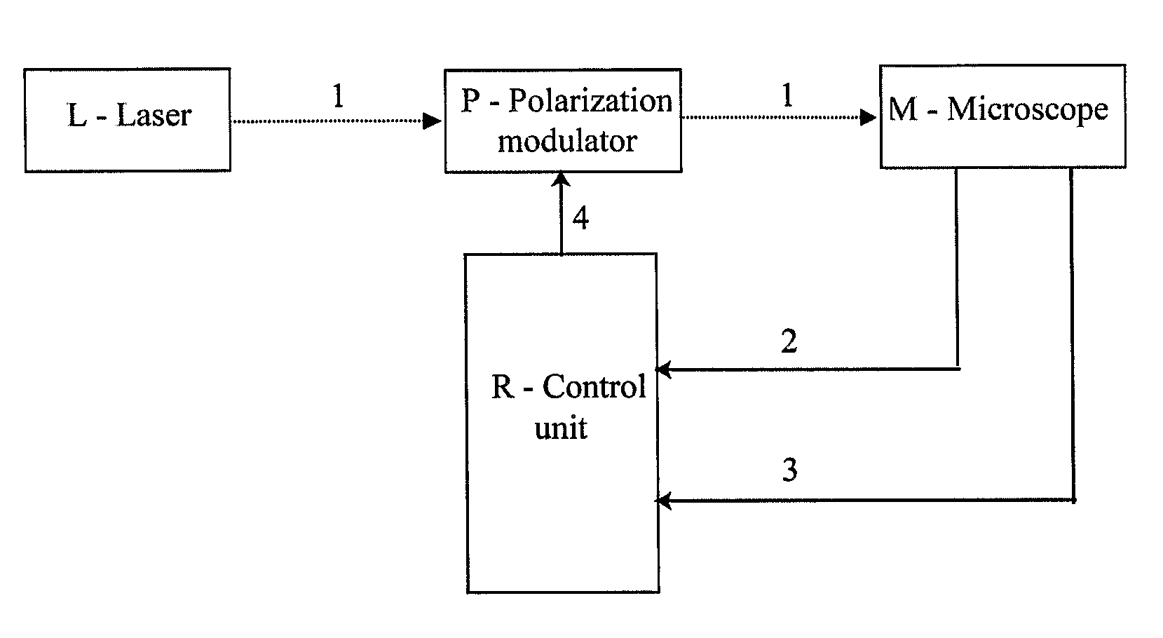
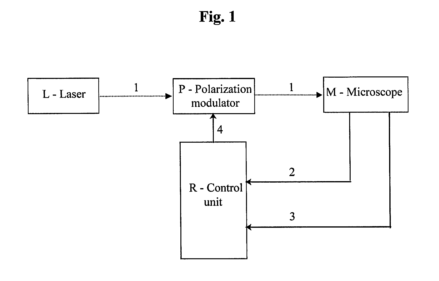
[0053]A device for carrying out polarization fluorescence microscopy, in principle illustrated in FIG. 1, has been created. The device comprises, as major components, a polarization modulator P acting on an excitation beam 1 emitted by a laser L, synchronized with a scanning fluorescence microscope M by a control unit R using timing signals 2 and 4.
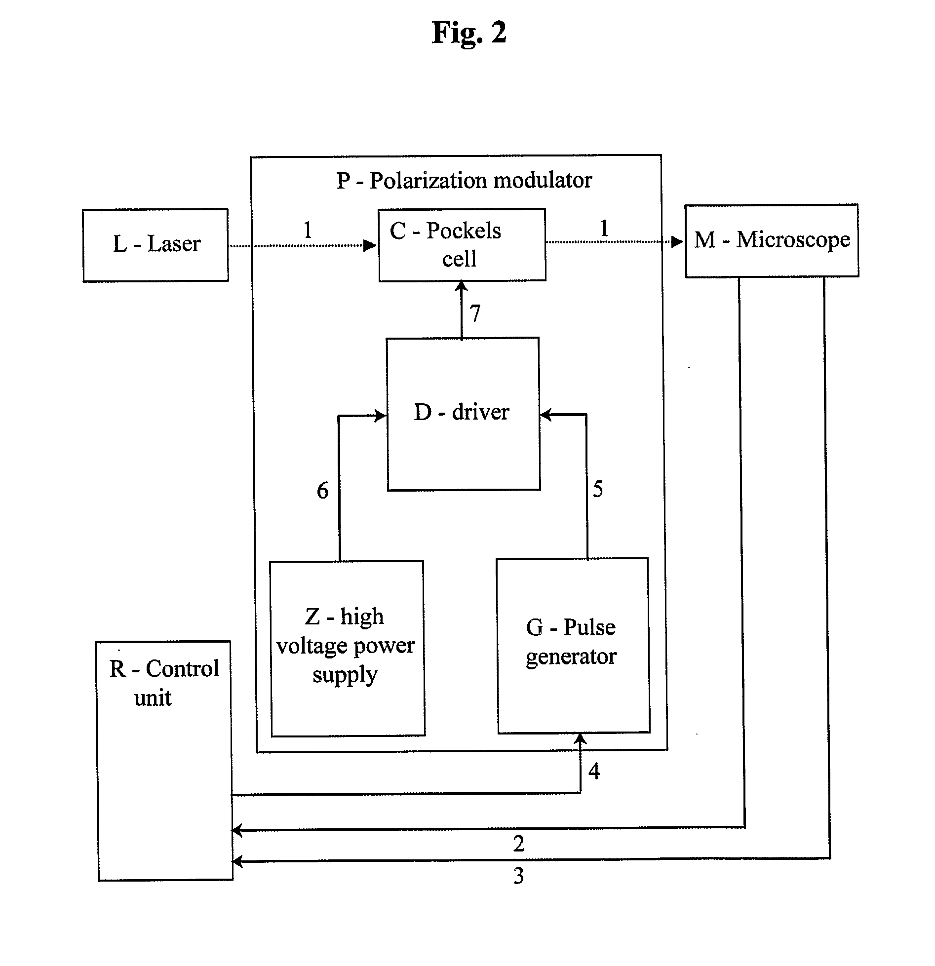
[0054]A device (shown in FIG. 2) has been designed to create a linearly polarized excitation laser beam 1 with direction of polarization modulated in synchrony with scanning of the microscope M. This was achieved by using a polarization modulator P consisting of a Pockels cell C (RTP-3-20-AR800-1000, Leysop Inc., United Kingdom) under the control of a high-voltage driver D (model B2, BME Bergmann GmbH, Germany). Driver D modulates high voltage 6 (0-1.2 kV), generated by a high voltage power supply Z (model HV, BME Berg...
example 2
Obtaining Structural and Functional Information About a Modified GFP Using Polarization Fluorescence Microscopy and the Device of Example 1
[0056]The device of Example 1 was used to acquire an image containing different parts (pixels) acquired with different polarizations of the excitation beam, i.e., a mixed image. The mixed image was deconvolved into separate individual images, each acquired with a different polarization of the excitation beam, in a manner schematically shown in FIG. 4. The result is illustrated in FIG. 5. Steps were taken during processing of the mixed image to account for delays in detector response and other factors. This was done by measuring the temporal response profile of the detectors used, and ascertaining what percentage of the signal (fluorescence) excited during a period of one polarization of the excitation beam is being reported in a pixel acquired during that period, and what percentage is being reported later. During deconvolution of the mixed image...
example 3
Monitoring and Determination of Protein Structure
[0064]The method and device for two-photon polarization microscopy of the invention have been used to determine and monitor structure of G-proteins, structure of a protein sensitive to calcium ion concentration, a protein sensitive to membrane voltage, receptor proteins, and a cytoskeleton attached protein (FIG. 6).
PUM
| Property | Measurement | Unit |
|---|---|---|
| time | aaaaa | aaaaa |
| voltage | aaaaa | aaaaa |
| polarization fluorescence microscopy | aaaaa | aaaaa |
Abstract
Description
Claims
Application Information
 - R&D
- Intellectual Property
- Life Sciences
- Materials
- Tech Scout
- Unparalleled Data Quality
- Higher Quality Content
- 60% Fewer Hallucinations
Browse by: Latest US Patents, China's latest patents, Technical Efficacy Thesaurus, Application Domain, Technology Topic, Popular Technical Reports.
© 2025 PatSnap. All rights reserved.Legal|Privacy policy|Modern Slavery Act Transparency Statement|Sitemap|About US| Contact US: help@patsnap.com


