
Techniques for accurately deriving physiologic parameters of a subject from photoplethysmographic measurements

a technology of physiologic parameters and photoplethysmography, which is applied in the field of medical devices and techniques for accurately deriving cardiac and breathing parameters of subjects, can solve the problems of inability particularly small mammals, or the current commercial pulse oximeters do not have the capability to accurately measure the heart rate of non-compliant subjects, and the pulse oximeter measurements are very susceptible to motion artifacts and breathing effort, so as roll improvement or reduction of undesirable frequency content conten

Active Publication Date: 2012-10-30
STARR LIFE SCI
View PDF55 Cites 14 Cited by
  • Summary
  • Abstract
  • Description
  • Claims
  • Application Information

AI Technical Summary

Benefits of technology

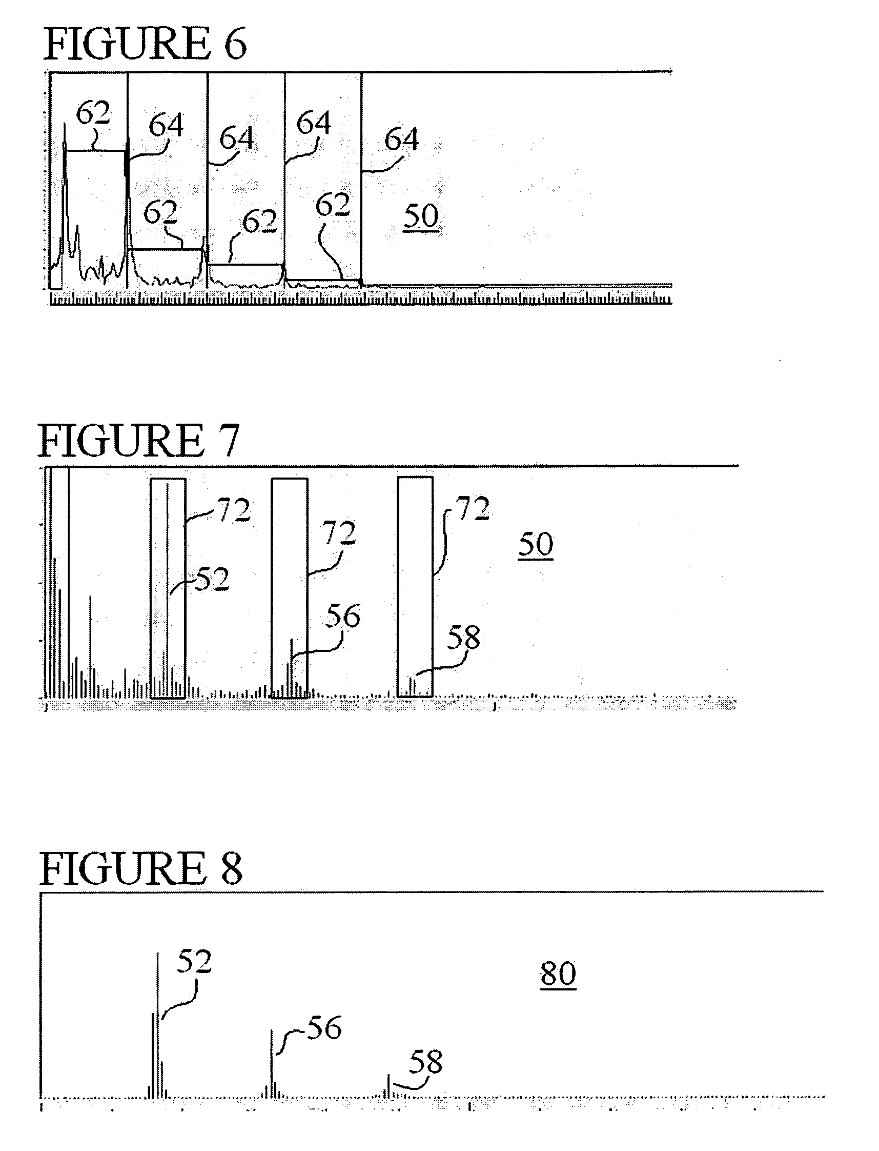
"The present invention provides techniques for isolating heart or breath rate data from a photoplethysmograph. These techniques involve filtering the frequency spectrum of the photoplethysmograph using a Fast Fourier Transform, detecting peaks in the frequency data set, and using an Inverse FFT to reconstruct the time-domain signal. The isolated heart or breath rate data can then be used to calculate physiologic parameters of the subject. The invention has several advantages, including improved accuracy and reliability in measuring physiologic parameters, and the ability to measure both heart rate and breath rate parameters simultaneously."

Problems solved by technology

Current commercial pulse oximeters do not have the capability to accurately measure heart rate of non-compliant subjects, particularly small mammals, or to accurately measure breath rate or other breathing related parameters in small subjects.
Pulse oximeter measurements are very susceptible to motion artifact.
One source of noise is breathing effort, which affects the blood volume delivered to the periphery in synchrony with the breathing cycle.
A more pernicious source of noise however, is caused by motion of the tissue and sensors.
Because the signal level of transmitted light is so small relative to the baseline transmitted light, very small motions of the tissue relative to the sensor pads can cause large changes in light transmission as the type and amount of tissue that resides between the LEDs and photodiode changes.
If these changes happen in a rhythmic manner, it can appear to the analysis software as a cardiac stroke volume pulse, or at the very least, it can swamp the ability of the software to pick out the heart rate signal.

Method used


Image

  • Techniques for accurately deriving physiologic parameters of a subject from photoplethysmographic measurements
  • Techniques for accurately deriving physiologic parameters of a subject from photoplethysmographic measurements
  • Techniques for accurately deriving physiologic parameters of a subject from photoplethysmographic measurements

Examples


Embodiment Construction

[0034]As described above, the present invention provides several techniques for isolating either heart or breath rate data from a photoplethysmograph, which is a time-domain signal or data set such as from known pulse oximeter sensors. A pulse oximeter system according to one aspect of the present invention is designed for small mammals such as mice and rats, such as using sensors sold under the Mouse OX™ brand by Starr Life Sciences, but the present invention is not limited to this application. The conventional pulse oximeters include a conventional light source, conventionally a pair of LED light sources, one being infrared and the other being red. The conventional pulse oximeters include a conventional receiver, typically a photo-diode. The light source and receiver are adapted to be attached to an external appendage of a subject, and may be secured to a spring-biased clip or other coupling device such as tape adhesives or the like. A specialized clip is available from Starr Life...


PUM


Abstract

Several techniques are disclosed for isolating either heart or breath rate data from a photoplethysmograph, which is a time domain signal such as from a pulse oximeter. The techniques involve the use of filtering in the frequency domain, after a Fast Fourier Transform (FFT) has been conducted on a given photoplethysmograph also references as a given set of discrete time-domain data. The filtering may be applied to an identified fundamental frequency and one or more harmonics for heart related parameters. The filter may be truncated to the frequency data set and further applied multiple times to improve roll off. After filtering, an Inverse FFT (IFFT) is used to reconstruct the time-domain signal, except with undesirable frequency content eliminated or reduced. Calculation or measurement of parameters is then conducted on this reconstructed time-domain signal.

Description

RELATED APPLICATIONS[0001]The present application claims the benefit of provisional patent application Ser. No. 60 / 884,389 entitled “Small Animal Pulse Oximeter Motion Artifact Rejection” filed Jan. 10, 2007.BACKGROUND OF THE INVENTION[0002]1. Field of the Invention[0003]The present invention relates to medical devices and techniques for accurately deriving cardiac and breathing parameters of a subject from extra-thoracic blood flow measurements, in particular, the invention relates to medical devices and techniques for deriving breath rate, breath distention, and pulse distention measurements of a subject from a pulse oximeter system coupled to a small animal.[0004]2. Background Information[0005]As background, one type of non-invasive physiologic sensor is a pulse monitor, also called a photoplethysmograph, which typically incorporates an incandescent lamp or light emitting diode (LED) to trans-illuminate an area of the subject, e.g. an appendage, which contains a sufficient amount...

Claims


Application Information

Patent Timeline
Patent Type & Authority Patents(United States)
IPC IPC(8): A61B5/0205
CPCA61B5/02405A61B5/02416A61B5/0816A61B5/7207A61B5/7253A61B5/7257A61B5/1455A61B5/0823
Inventor HETE, BERNARD F.AYERS, ERIC J
Owner STARR LIFE SCI
Learn More