
Phage display technologies

a technology of phages and coat proteins, applied in the field of improved phagemid vectors, can solve the problems of large size and cloning strategy limitations of heterologous sequences, affecting the efficiency of the system, and affecting the choice of coat proteins for display

Active Publication Date: 2010-10-12
RIBOVAX BIOTECHNOLOGIES SA
View PDF1 Cites 6 Cited by
  • Summary
  • Abstract
  • Description
  • Claims
  • Application Information

AI Technical Summary

Benefits of technology

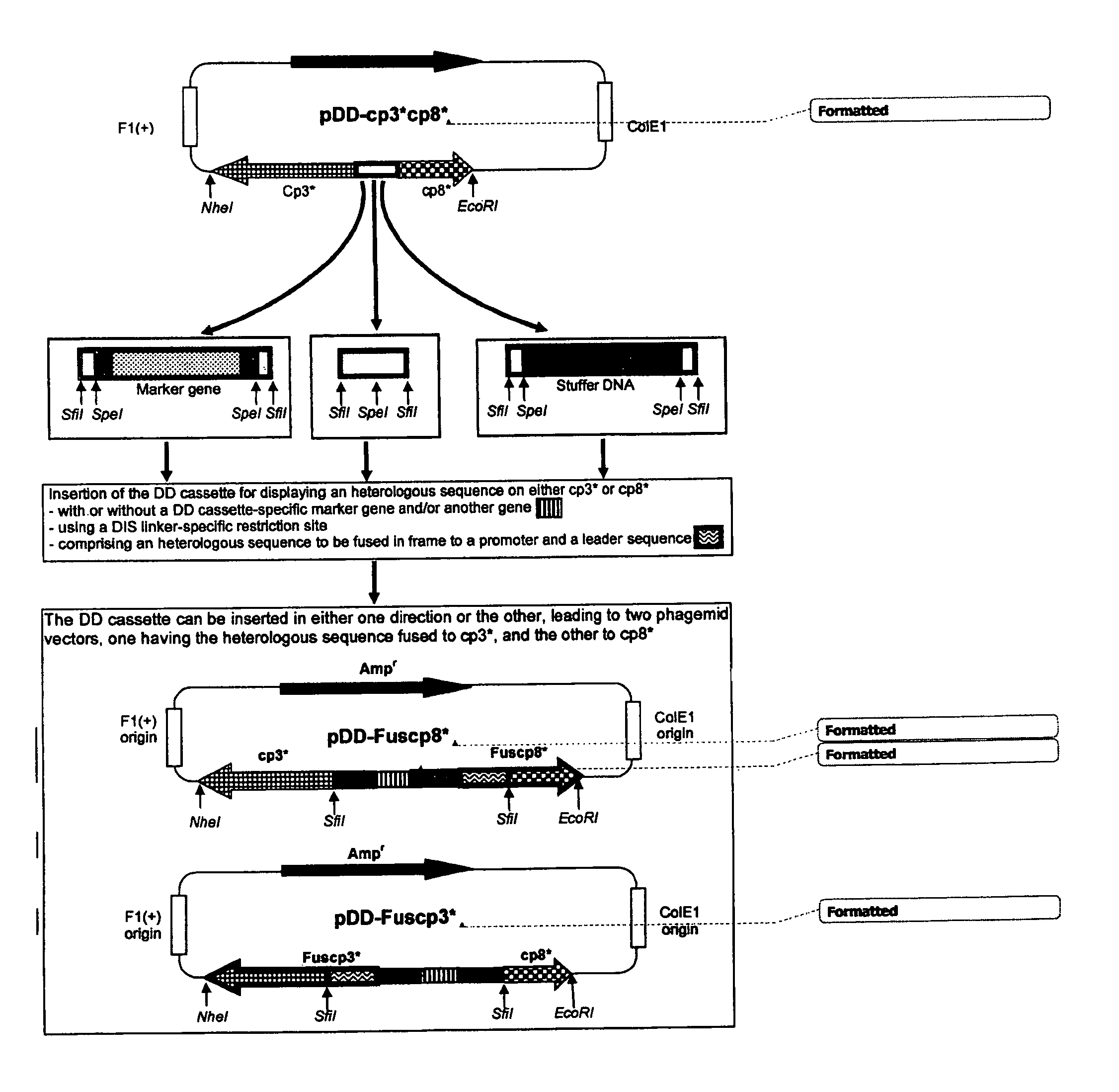
"The present invention provides a method for cloning, producing, and screening amino acid sequences fused to either one or the other of two functional coat proteins on the surface of filamentous phage using a single phagemid vector and a single cloning and transformation step. This allows for the bidirectional cloning of a DNA coding for an amino acid sequence to be fused at the N-terminus of either one or the other of two functional coat proteins. The invention also provides phage or cell libraries containing fused protein sequences fused at the N-terminus of either one or the other of two functional coat proteins by means of aDIS linker. The use of these vectors and methods facilitates the production of phage or cell libraries with improved efficiency and accuracy."

Problems solved by technology

When using a phage vector, the DNA coding for the fusion protein is directly cloned into a coat protein of the phage genome, allowing high display levels but with a strong limitation in the size and in the cloning strategy of the heterologous sequence.
While very efficient, the phage display technologies currently available still have room for improvement, and the choice of the coat protein for the display still remains an important issue.
In fact, the efficiency of the system can be substantially affected by the “fitness” of the protein sequence to be expressed, displayed, and screened using one rather than another specific coat protein sequence, a property that cannot be possible to determine in advance.
However, given that the affinity of the antibodies in a library is highly variable and impossible to foresee, other factors can affect the display system such as the proteolytic degradation of the cp3- / cp8-based fusion protein in the periplasmic space of E. coli or recombination events eliminating sequences included in the phagemid vector.

Method used


Image

  • Phage display technologies
  • Phage display technologies
  • Phage display technologies

Examples


example 1

Preparation and Expression of a DIS Linker2-Modified DNA Sequence Encoding a Filamentous Phage Coat Protein 3 (cp3*) for Displaying Proteins and Peptides

[0152]a) Materials & Methods

[0153]Production of M13 Helper Double-Stranded DNA.

[0154]A commercially available bacteriophage M13 helper (VCSM13, Stratagene, La Jolla, Calif.) was used as a source for isolating its double-stranded replicative form DNA. Into 2 ml of LB medium, 50 μl of a culture of a bacterial strain carrying an F′ episome (E. Coli XL1 Blue, Stratagene, La Jolla, Calif.) was admixed with 1×10¹¹ bacteriophage particles. The admixture was incubated for 4 to 5 hours at 37° C. with constant agitation. The admixture was then centrifuged at 12,000×g for 5 minutes to pellet the infected bacteria. After the supernatant was removed, the bacteria pellet was used in a standard DNA extraction protocol using Qiagen mini prep kit and analysed in 1% agarose gel electrophoresis.

[0155]Production of a Gene Encoding a DIS Linker2 N-Termi...

example 2

Preparation and Expression of a DIS Linker2-Modified DNA Sequence Encoding a Filamentous Phage Coat Protein 8 (cp8*) for Displaying Proteins and Peptides

[0176]a) Materials & Methods

[0177]Production of a Gene Encoding a DIS Linker2 N-Terminal Modified cp8 Protein (cp8* Protein), Related Vectors Expressing cp8*-Based Fusion Proteins, and their Analysis

[0178]The production of cp8* starting from a commercially available bacteriophage M13 helper and the cloning strategy are identical to those indicated in Example 1 for cp3*. The PCR was carried out using the primers cp8*FW and cp8*RW (SEQ ID NO.: 15 and SEQ ID NO.: 16; Table I). The PCR conditions used were: 95° C. for 2 min (1 cycle); 95° C. for 20 sec, 63° C. for 30 sec, 72° C. for 40 sec (35 cycles). The PCR amplification was carried out using Expand High Fidelity (Roche) according to the manual instructions. The DNA template (obtained from VCSM13) and the primers concentration used were the same described for the cp3* PCR amplificati...

example 3

Functional Validation of Phage Displaying Cp3* or Cp8* Fused to a Peptide or an Antibody

[0186]a) Materials & Methods

[0187]Amplification of the Phagemids

[0188]The following methods were performed as described in the literature (“Molecular Cloning: A Laboratory Manual”, Sambrook et al., Cold Spring Harbor Press, NY, 1989). Electrocompetent E. coli XL1-Blue cells were electroporated (0.2 cm E. coli Pulser cuvette, 2.5 Kv) with 50 ng of the phagemid vector (pRIB1-cp3, pRIB-HAcp3, pRIB-e44 cp3, pRIB1-cp3*, pRIB1-HAcp3*, pRIB1-e44 cp3*, pRIB2-cp8*, pRIB2-HAcp8*, or pRIB2-e44 cp8*).

[0189]Each cuvette was flushed immediately with 1 ml and then with 2 ml of SOC medium (Sodium chloride 0.5 g / L, Tryptone 20.0 g / L, Yeast extract 5.0 g / L, KCl 2.5 mMol, pH adjusted to 7.0 with NaOH: after autoclaving, 5 ml of a 2 M MgCl2 sterile solution and 20 ml of a 1 M sterile glucose solution for 1 liter of medium are added just before use) at room temperature. The 3 ml culture was transferred into a 50 ml p...


PUM

PropertyMeasurementUnit
temperatureaaaaaaaaaa
concentrationaaaaaaaaaa

Abstract

The present invention provides novel technologies for producing and screening fusion proteins on the surface of filamentous phage. In particular, a single vector can be used for generating cell and phage libraries containing a given series of protein sequences fused to either one or other of two phage coat proteins. This approach simplifies and improves the efficiency of the subsequent phage display-based selection of protein-binding molecules having therapeutic or diagnostic utility, such as antibodies, peptides, or epitope-binding regions.

Description

[0001]This application is a National Stage entry of International Application No. PCT / IB2006 / 001878, filed Jul. 6, 2006, the entire specification claims and drawings of which are incorporated herewith by reference.FIELD OF THE INVENTION[0002]The invention relates to improved phagemid vectors for generating phage display libraries.BACKGROUND OF THE INVENTION[0003]Phage display-based technologies provide means for cloning, expressing, selecting, and engineering polypeptides with biological functions mediated by their binding to another protein or any other biological target. The iterative process of the affinity-based selection allows the enrichment into relevant clones isolated from large libraries of protein sequences, such as antibodies, epitopes, antigens, bioactive peptides, enzyme inhibitors, enzymes, DNA-binding proteins, isolated protein domains, or ligands for receptors.[0004]In combination with other techniques, phage display technologies, starting from sequences isolated fr...

Claims


Application Information

Patent Timeline
Patent Type & Authority Patents(United States)
IPC IPC(8): C40B40/02C40B50/06C12N15/87A61K39/00
CPCC12N15/1037C12N15/64C12N15/09C12N15/10
Inventor BURIONI, ROBERTOCLEMENTI, MASSIMO
Owner RIBOVAX BIOTECHNOLOGIES SA
Learn More