Diagnosis of liver pathology through assessment of protein glycosylation
a technology of protein glycosylation and liver disease, applied in the field of immunodeficiency diagnosis, can solve the problems of liver failure, liver cirrhosis is a leading cause of death among adults, and many liver diseases, including hbv and hcv infection, can be asymptomatic for many
- Summary
- Abstract
- Description
- Claims
- Application Information
AI Technical Summary
Benefits of technology
Problems solved by technology
Method used
Image
Examples
example 1
General Experimental Procedures
[0071]Glycan Analysis. Serum was obtained from patients clinically diagnosed as infected with chronic HBV, and from patients clinically diagnosed as having HCC, patients clinically diagnosed as having liver cirrhosis, and from control subjects with no evidence of any liver disease. Serum samples were stored at −80° C. until analysis.
[0072]Protein aliquots (1 mg / mL) were denatured with 1% SDS, 50 mM β-mercaptoethanol for 10 min at 100° C. The solution was cooled, and supplemented with NP-40 to a concentration of 5.75%. PNGase F (ProZyme, San Leandro, Calif.) was added to a final concentration of 1 mU(IUB) / μL, and a cocktail of protease inhibitors was added to the mix. The solution was then incubated for 24 hours at 37
[0073]° C. The oligosaccharides separated from the proteins in the sample were recovered and purified by solid phase extraction using a porous graphite matrix (LudgerClean H, Ludger Limited, Oxford, UK). Free oligosaccharides were labeled w...
example 2
Core Fucosylation is Increased in Hepatocellular Carcinoma
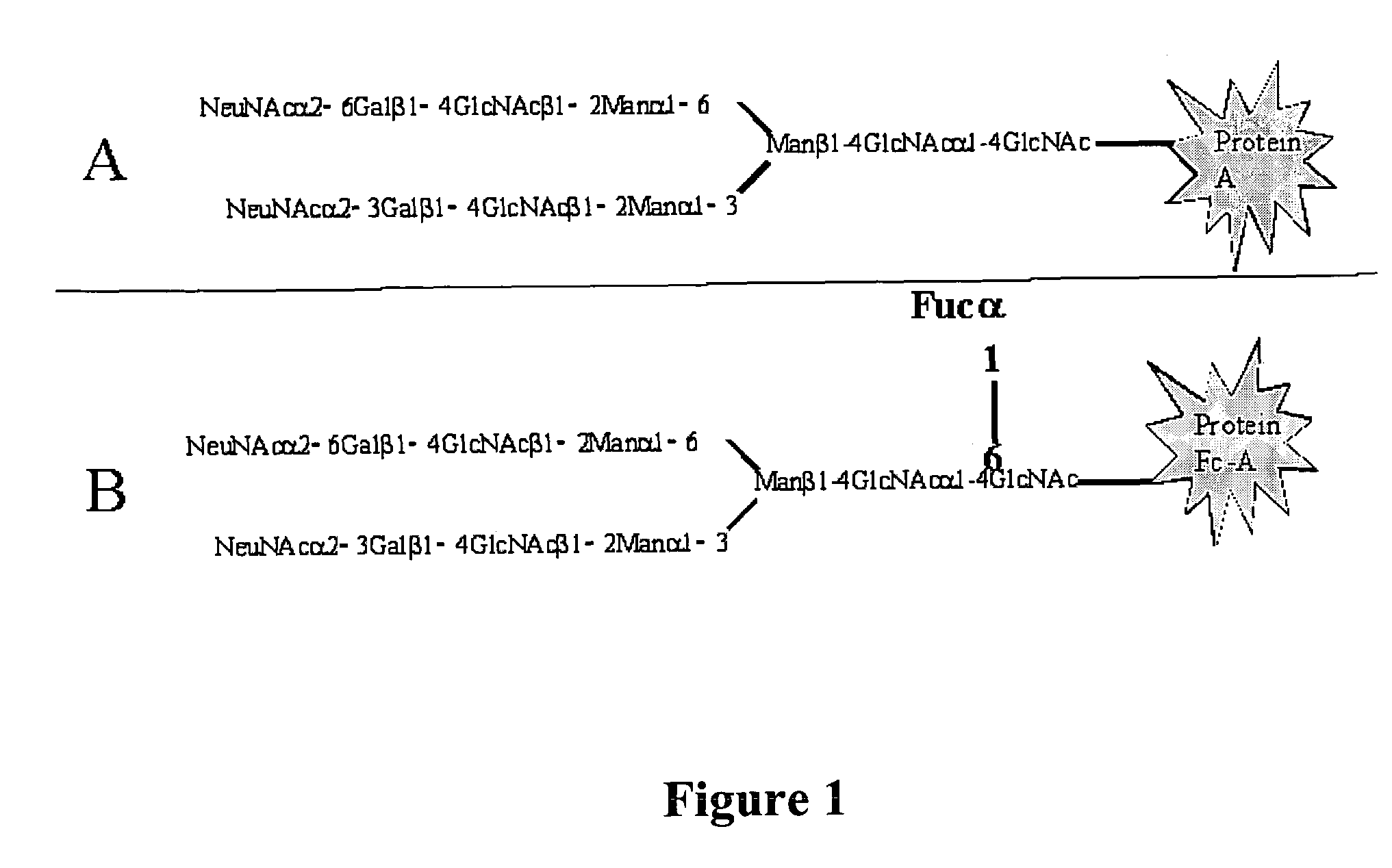
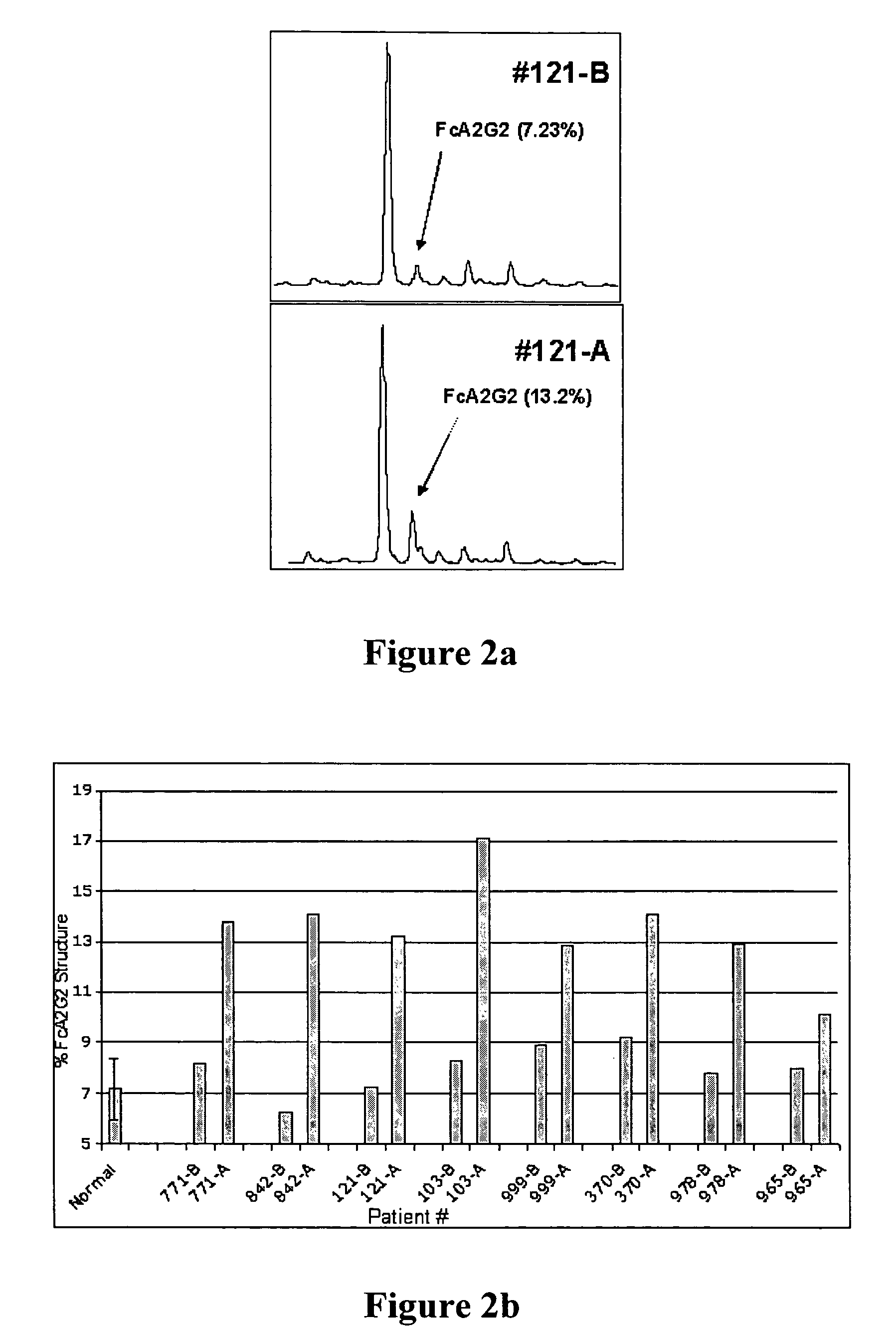
[0082]A targeted glycoproteomic methodology that allows for the identification of glycoprotein biomarkers in serum was developed. The methodology first identifies changes in N-linked glycosylation that occur with the disease. These changes act as a tag so that specific proteins that contain that glycan structure can be extracted. Initial experiments in an animal model revealed a protein, GP73, that is more sensitive at detecting HCC than currently used markers such as AFP. In the animal model of HCC, the change in glycosylation was an increase in core fucosylation (Block et al. 2005). This change was also observed in people who developed HCC. An example of the increase in fucosylation is shown in FIG. 2. This sample set consisted of 16 samples from 8 patients at two time points: before the development of cancer, and after the diagnosis of cancer. In this case, all patients classified as before were clinically diagnosed with c...
example 3
Identification of Fucosylated Glycoproteins in Hepatocellular Carcinoma
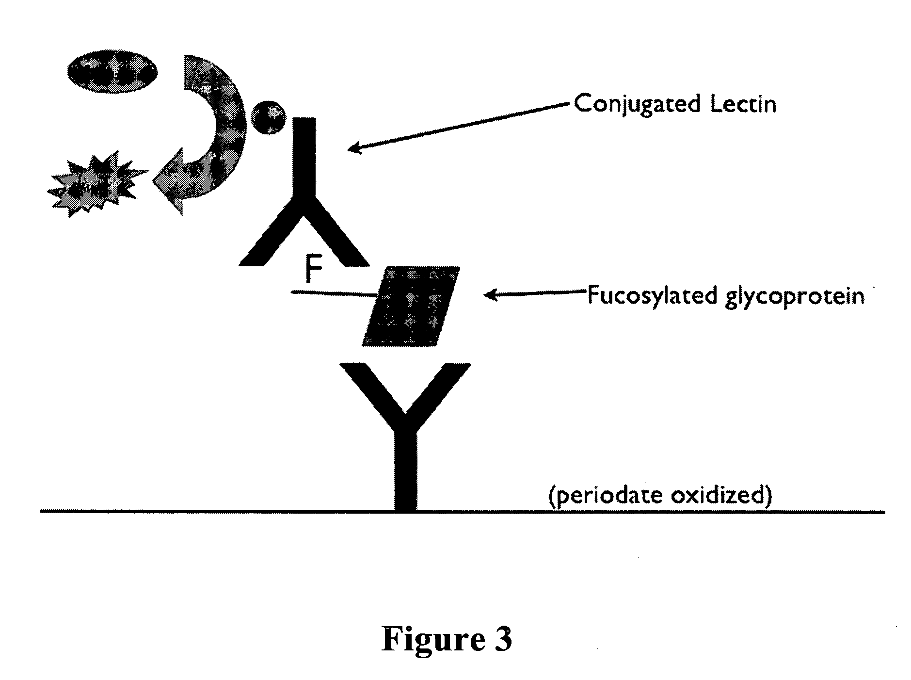
[0083]As the level of fucosylation was increased in patients with HCC, it was imperative to determine those proteins that had increased fucosylation. Therefore, the fucosylated glycoproteins associated with sera from either pooled normal or pooled HCC positive individuals were extracted using lectins and the proteome analyzed by either two dimensional gel electrophoresis (2DE) or by a simple LC MS / MS based methodology. Using these methodologies it was observed that glycoproteins such as fucosylated (Fc) α-1-acid glycoprotein, Fc-ceruloplasmin, Fc-α-2-macroglobulin, Fc-hemopexin, Fc-Apo-D, Fc-HBsAg, and Fc-Kininogen increased in patients with HCC, while the levels of Fc-haptoglobin decrease in the those patients (Table 1). Interestingly, several immunoglobulin molecules (IgG, IgA and IgM) were also found in the fucosylated proteome of those patients with cancer. However, further analysis has shown that these prote...
PUM
 Abstract
Description
Claims
Application Information
 - R&D
- Intellectual Property
- Life Sciences
- Materials
- Tech Scout
- Unparalleled Data Quality
- Higher Quality Content
- 60% Fewer Hallucinations
Browse by: Latest US Patents, China's latest patents, Technical Efficacy Thesaurus, Application Domain, Technology Topic, Popular Technical Reports.
© 2025 PatSnap. All rights reserved.Legal|Privacy policy|Modern Slavery Act Transparency Statement|Sitemap|About US| Contact US: help@patsnap.com


