Typically, the heart loses propulsive power because the
cardiac muscle loses capacity to stretch and contract.
Often, the ventricles do not adequately eject or fill with blood between heartbeats and the valves regulating
blood flow become leaky, allowing regurgitation or back-flow of blood.
The impairment of arterial circulation deprives vital organs of
oxygen and nutrients.
Fatigue,
weakness and the inability to carry out daily tasks may result.
As
heart failure progresses, it tends to become increasingly difficult to manage.
Even the compensatory responses it triggers in the body may themselves eventually complicate the
clinical prognosis.
If the
oxygen supply falls short of the growing demand, as it often does, further injury to the heart may result.
A particularly severe form of
heart failure is congestive heart failure (CHF) wherein the weak pumping of the heart leads to build-up of fluids in the lungs and other organs and tissues.
Briefly, the poor cardiac function resulting from heart failure can cause blood to back up in the lungs, thereby increasing
blood pressure in the lungs.
This can cause severe respiratory problems and, left untreated, can be fatal.
In this regard, previous techniques for detecting pulmonary
edema based on impedance are often unduly complex.
In some cases, changes in impedance due to other factors besides the fluids associated with pulmonary
edema can possibly result in a false positive detection of pulmonary
edema.
With CSA, the nerve signals are not properly generated during extended periods of time while the patient is asleep or are of insufficient magnitude to trigger sufficient
muscle contraction to achieve
inhalation.
Arousal from sleep due to CSA usually lasts only a few seconds, but such brief arousals nevertheless disrupt continuous sleep and can prevent the patient from achieving rapid
eye movement (REM) sleep, which is needed.
Due to poor cardiac function caused by CHF, patients already suffer from generally low blood
oxygen levels.
Frequent periods of
sleep apnea result in even lower blood oxygen levels.
However, an obstruction of the respiration
airway blocks delivery of air to the lungs and so blood CO2 levels continue to increase, usually until the patient awakens and readjusts his or her position so as to reopen the obstructed respiration pathway so that normal
breathing can resume.
However, while asleep, the muscles can relax to a point where the
airway collapses and hence becomes obstructed.
As with CSA,
arousal from sleep usually lasts only a few seconds but is sufficient to disrupt continuous sleep and prevent proper REM sleep.
In addition, patients are at greater risk of OSA with increasing age, due to loss of
muscle mass, particularly within the muscles that would otherwise hold the respiration
airway open.
With CHF, poor cardiac function results in poor
blood flow to the brain such that
respiratory control nerve centers respond to blood CO2 levels that are no longer properly representative of the overall blood CO2 levels in the body.
This cycle becomes increasingly unbalanced until respiration alternates between
hypopnea and hyperpnea.
Problems, however, can arise when attempting to detect
apnea /
hypopnea via thoracic impedance in patients with pulmonary edema.
The present inventor has noted that apnea detection via thoracic impedance is most reliable when the lungs are clear but is considerably less reliable when the lungs are filled with fluids, i.e. when the patient suffers from pulmonary edema.
Hence, it is not always possible to reliably detect apnea using an impedance-based detection technique if the patient suffers from pulmonary edema.
However, if the
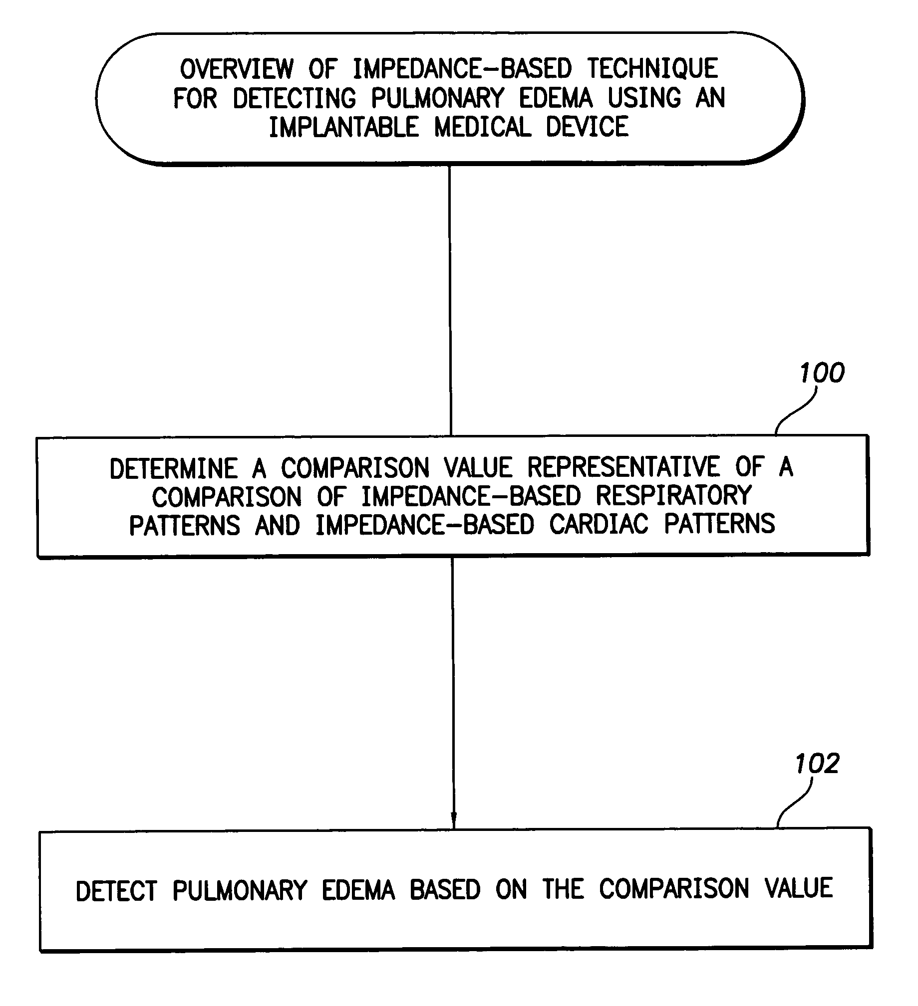
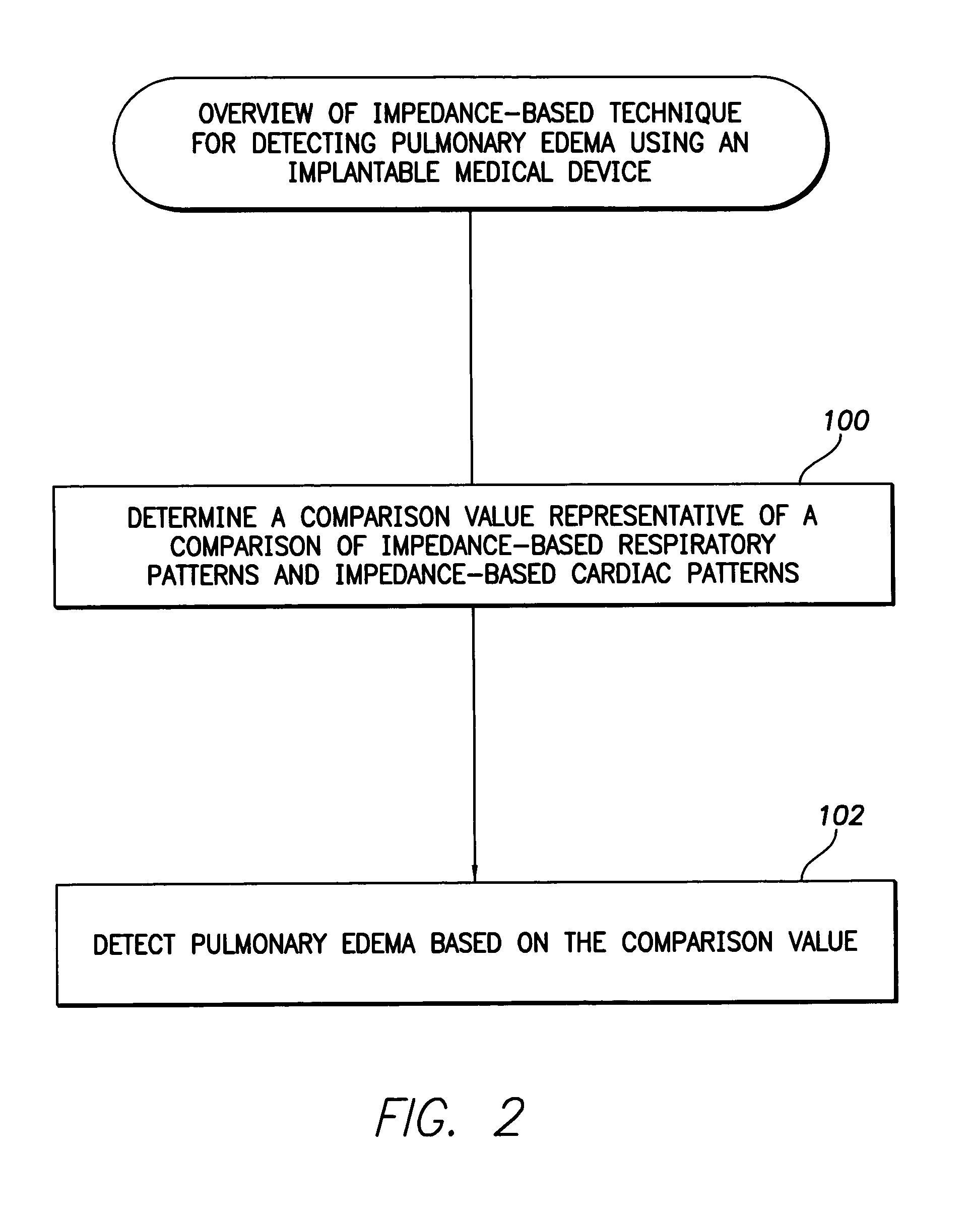
amplitude ratio falls below the threshold, this is an indication that pulmonary edema has occurred within the patient and the lungs are filled with fluids.
Indeed, during pulmonary edema,
respiratory effort often increases as the patient attempts to breathe more deeply to compensate for the reduction in oxygen entering the
blood stream due to the fluidic
lung.
In many cases, pulmonary edema is associated with heart failure.
However, if the ratio falls below the threshold, this is an indication that impedance-based techniques for detecting episodes of reduced respiration can no longer reliably be performed because variations in the impedance signals due to respiration are too severely diminished relative to those due to the beating of the heart.
In other words, respiration patterns can no longer be reliably derived by
low pass filtering impedance signals and hence a detector that seeks to detect episodes of reduced respiration based on an analysis of impedance signals can no longer be deemed reliable.


