
Nanoarrays of single virus particles, methods and instrumentation for the fabrication and use thereof

a technology of single virus particles and nano-arrays, which is applied in the field of nano-arrays of single virus particles, methods and instruments for the fabrication and use thereof, can solve the problems of limited practical resolution of current microarray technologies, such as spotting with pin arrays, inkjet printing or methods derived from photolithography, and the density of fabricated arrays, and the number of distinct deposited biological entities (including but not limited to proteins, nucleic acids, carbohydrates,

Active Publication Date: 2009-08-04
NORTHWESTERN UNIV
View PDF5 Cites 13 Cited by
  • Summary
  • Abstract
  • Description
  • Claims
  • Application Information

AI Technical Summary

Benefits of technology

The invention is about creating arrays of biological entities, specifically virus particles, on surfaces using a nanolithographic printing process. The invention also includes methods for making these arrays and using them for various applications. The technical effects of the invention include the ability to create highly organized and controlled arrays of virus particles on surfaces, which can be useful for various research and development purposes.

Problems solved by technology

[5] However, current microarraying technologies, such as spotting with pin arrays, inkjet printing or methods derived from photolithography, are limited in their practical resolution—from hundreds to tens of microns, depending on the technique.
The density of the fabricated arrays and therefore the number of distinct deposited biological entities (which include but are not limited to proteins, nucleic acids, carbohydrates, lipids, and especially complex or assemblies thereof, such as viruses or cell components) are therefore limited.
This method offers little if any control over the density and placement of said particles.
This is in part because of limited resolution (vide supra), the size of the particles interrogated, and especially the chemistry used to immobilize them.
Indeed, prior efforts have focused on the genetic modification of a virus particle to present unnatural surface binding functionality to the patterned interface.

Method used


Image

  • Nanoarrays of single virus particles, methods and instrumentation for the fabrication and use thereof
  • Nanoarrays of single virus particles, methods and instrumentation for the fabrication and use thereof
  • Nanoarrays of single virus particles, methods and instrumentation for the fabrication and use thereof

Examples


working examples

[0085]Non-limiting working examples are provided.

working example

Site-Isolated TMV Arrays

[0086]In this example, virus particles were site-isolated, positioned and oriented on Zn2+-MHA nanotemplates generated by dip pen nanolithography. Viral immobilization was characterized using antibody-virus recognition as well as infrared spectroscopy.

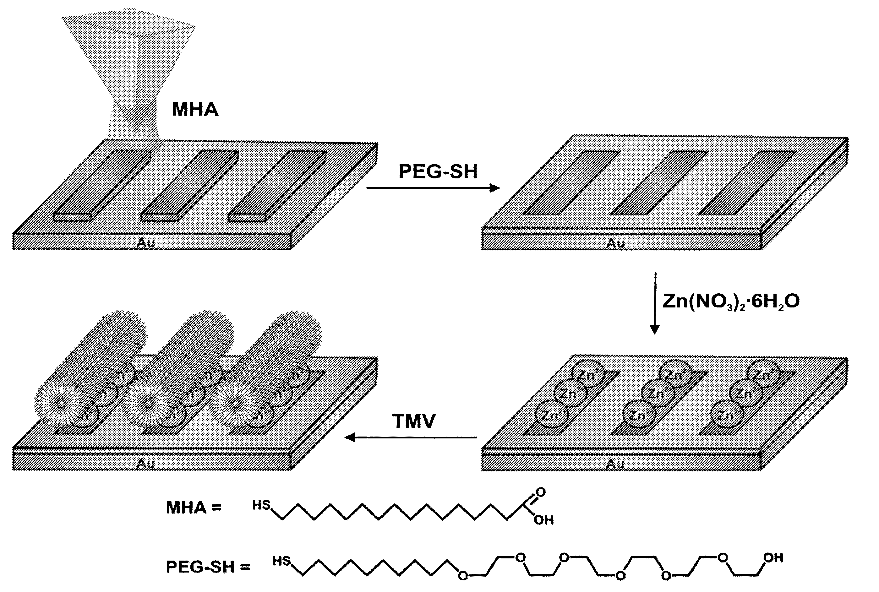
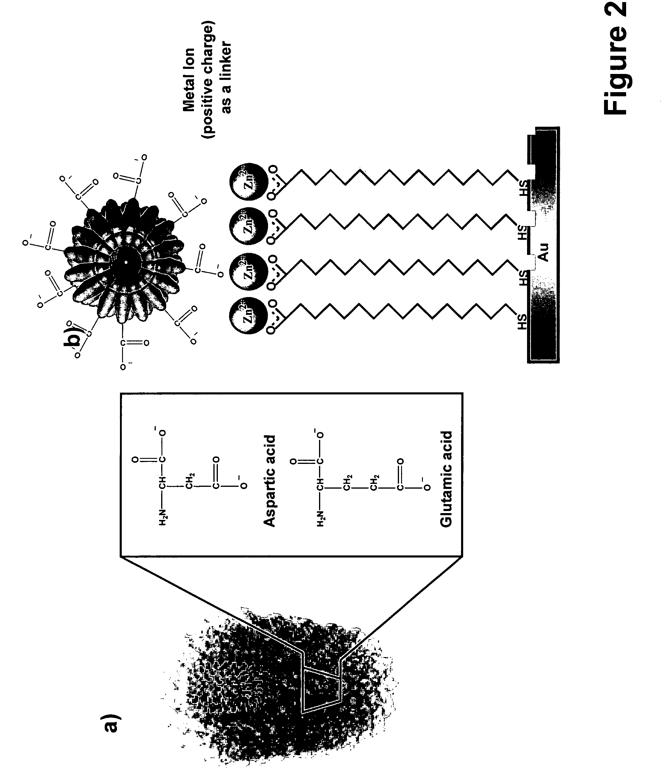
[0087]The approach used in this working example relied on the ability of metal ions (Zn2+) to bridge a surface patterned with features made of 16-mercaptohexadecanoic acid (MHA) and the TMV with its carboxylate-rich surface.

[0088]Tobacco Mosaic Virus (TMV) was chosen because of its anisotropic tubular structure (about 300 nm long, 18 nm diameter), size, stability, and well -characterized carboxylate-rich surface.[10] It serves as an excellent demonstrative system to evaluate how one can use DPN to control the positioning and orientation of nanoscale virus particles within an extended array.

[0089]Virus nanoarrays were fabricated by initially generating chemical templates of MHA on a gold thin film using DPN (FIG....


PUM

PropertyMeasurementUnit
surface areaaaaaaaaaaa

Abstract

A nanoarray template utilizing coordination chemistry or metal ion binding to control the site-isolation and orientation of virus particles is provided. The nanoarray template is generated by lithography including Dip Pen Nanolithography. The surface chemistry that is inherent in many viruses, metal-ion based or inorganic coordination chemistry is used to immobilize individual virus particles without the need for their genetic modification. Single particle control enables a wide variety of studies involving viruses that are not possible with microarrays, including single particle, single cell infectivity studies, exploration of such structures as templates in materials synthesis and molecular electronics, and studies aimed at understanding how surface presentation influences their bioactivity. This is an example of such control at the single-particle level, and therefore, commercial use of nanoarrays in biological systems.

Description

RELATED APPLICATIONS[0001]This application claims priority benefit to provisional application Ser. No. 60 / 712,432 filed Aug. 31, 2005 to Mirkin, et al., which is hereby incorporated by reference in its entirety.GOVERNMENT RIGHTS[0002]This invention was made with government support under following grants: Air Force Office of Science Research (AFOSR) grant MURI F49620-00-1-0283; NIH grant 1DP1OD000285-01; NSF / NSEC grant EEC-0118025; DARPA / AFOSR grant FA9550-05-1-0348; and ARO-MURI 28065-3-A1 / / W91NF-04-1-071. The government has rights in this invention.BACKGROUND[0003]Microarray technology has led to significant advances in many areas of medical and biological research,[1] opening up avenues for the combinatorial screening and identification of single nucleotide polymorphisms (SNPs),[2] high sensitivity expression profiling of proteins,[3,4] and high throughput analysis of protein function.[5] However, current microarraying technologies, such as spotting with pin arrays, inkjet printin...

Claims


Application Information

Patent Timeline
Patent Type & Authority Patents(United States)
IPC IPC(8): C12Q1/70C12N7/00C12P1/00C12N11/00
CPCA61K31/70B82Y5/00G01N33/56983C12N7/00B82Y30/00C12N2770/00011Y10S977/802G01N33/50G01N33/53G01N33/569
Inventor MIRKIN, CHAD A.VEGA, RAFAEL A.MASPOCH, DANIELSALAITA, KHALID
Owner NORTHWESTERN UNIV
Learn More