Ophthalmic microscope

a microscope and ophthalmology technology, applied in the field of ophthalmology microscopes, can solve the problems of reduced visibility, unfavorable fusion of images, reduced visual performance, etc., and achieve the effects of reducing price, widening the observation range of the inspecting eye, and changing quickly

Inactive Publication Date: 2008-10-28
KK TOPCON
View PDF5 Cites 5 Cited by
  • Summary
  • Abstract
  • Description
  • Claims
  • Application Information

AI Technical Summary

Benefits of technology

[0016]According to the characteristics of the present invention, since the correlated color temperature changing means enables the changing of the correlated color temperature of the illumination light, operating in conjunction with the switching of the injection angle of the illumination light, the correlated color temperature of the light source can be readily changed while simultaneously changing the injection angle of the illumination light. Therefore, it becomes possible to set the observation conditions according to the medical condition of the inspecting eye and expand the observation range of the inspecting eye. In addition, since the microscope is multi-functional, reduction of the price is not necessary and excessive storage space is not required, allowing the device to be used in hospitals with limited space or the like.

Problems solved by technology

“Cataracts” is an ophthalmic disease, wherein the natural lens of the eye becomes clouded due to aging or other causes, leading to decreased visual performance.
However, in the case of 0° lighting, since the red reflex in the observation beam varies from right to left, there are problems in that, for example, the image is not favorably fused when viewing with binocular vision.
Thus, there is a problem of reduced visibility.
However, as in the case of cataracts, when the degree of cloudiness (intensity of the reflected light) varies depending on the progression of the condition where cloudiness spreads over the lens, for example, in the case of hypermature cataracts, a red reflex cannot be obtained even with complete coaxis lighting.
It is extremely difficult to perform CCC (Continuous Circular Capsulorrhoxis, i.e. incision of the capsule) without a red reflex.
However, since the capsule of the lens has extremely high transparency and low reflection intensity, it is difficult to obtain sufficient visibility.
Therefore, defects occur in microscopes employing a xenon light source.
However, it is difficult to say that the above problems can be completely prevented, since a changing operation or the like is still required.

Method used


Image

  • Ophthalmic microscope
  • Ophthalmic microscope
  • Ophthalmic microscope

Examples


embodiment 1

(Structure of Each Component and the Entirety of the Ophthalmic Microscope)

[0033]Embodiment 1 of the present invention is an ophthalmic microscope equipped with a filter, which is a component of the correlated color temperature changing means, for changing the correlated color temperature, wherein the correlated color temperature changing means and the lighting angle switching means are constituted together by employing a closure plate containing a transparent piece that is a component of the lighting angle switching means.

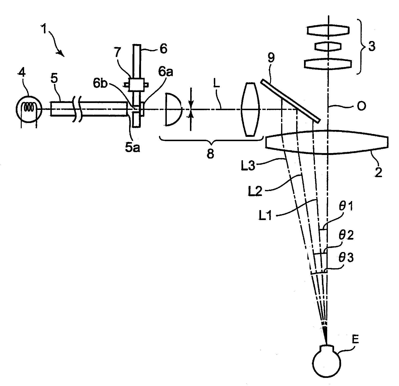
[0034]FIG. 1 shows an outlined structure of the ophthalmic microscope 1 in the embodiment of the present invention. The ophthalmic microscope 1 enables observation through binocular vision. For example, it is placed on the objective lens 2 opposite to the inspecting eye E and the extension of the optical axis of the objective lens 2. The ophthalmic microscope 1 is equipped with an ocular lens component comprising an ocular lens for the left and right eyes (not sho...

embodiment 2

[0056]In Embodiment 1, an ophthalmic microscope equipped with a halogen light source was explained for the purpose of example. It is also possible to place a xenon light source in the ophthalmic microscope of said Embodiment 1 and Embodiments 3 and 4 mentioned below. Furthermore, since the components are almost the same except for the xenon light source and the closure plate 61 shown in FIG. 7, only different components will be explained to clarify the difference from Embodiment 1.

[0057]When using the ophthalmic microscope equipped with a xenon light source, the correlated color temperature changing filter is used to change the correlated color temperature of the illumination light from 6000K to 3000K. The closure plate 61 formed in a disc shape, as shown in FIG. 7 for example, is used as the correlated color temperature changing means. Similar to the closure plate 6 (see FIG. 3A), the transparent pieces 61b1 to 61b4 are open near the periphery of the closure plate 61. The transpare...

embodiment 3

(Structure of Each Part and the Entire Ophthalmic Microscope)

[0060]Embodiment 3 of the present invention is an ophthalmic microscope comprising a closure plate containing a correlated color temperature changing filter which is a component of the correlated color temperature changing means, and a transparent piece which is a component of the lighting angle switching means, different from the constitution of the microscope of Embodiment 1, wherein the color temperature changing means and the lighting angle switching means are separately constituted.

[0061]The difference from the ophthalmic microscope of Embodiment 1 is that the ophthalmic microscope comprises the closure plate 6 and said correlated color temperature changing filter 6a shown in FIG. 3A as individual components, wherein the closure plate 6 works in conjunction with the correlated color temperature changing filter 6a to drive. The closure plate 6 used in the explanation, i.e., the closure plate 6 on which the correlated c...


PUM


Abstract

An ophthalmic microscope including an objective lens placed in face of an inspecting eye; a light source for radiating an illumination light, an illumination optics for directing the illumination light to the inspecting eye via the objective lens, an observation optics placed along with an optical axis of the objective lens, a lighting angle switch placed on a light path of the illumination light for changing the angle of incidence of the illumination light to the inspecting eye, and a correlated color temperature changer working in conjunction with the switching of the angle of incidence by the lighting angle switch to change the correlated color temperature of the illumination light.

Description

BACKGROUND OF THE INVENTION[0001]1. Field of the Invention[0002]The present invention relates to an ophthalmic microscope.[0003]2. Description of the Related Art[0004]Various ophthalmic operations have been conventionally performed. In particular, cataract surgery is one example of an ophthalmic operation that has been typically performed. “Cataracts” is an ophthalmic disease, wherein the natural lens of the eye becomes clouded due to aging or other causes, leading to decreased visual performance. A procedure called phacoemulsification (hereinafter, referred to as “the suction method”) has been a commonly performed operation for cataracts. In the suction method, an incision is made along the edge of the cornea and the capsule, an aspirator is inserted into the incision to remove the clouded lens, and an intraocular lens (IOL) is implanted in place of the removed lens.[0005]In the suction method, a surgical microscope is used to obtain an enlarged image of the patient's eye (hereinaf...

Claims


Application Information

Patent Timeline
Patent Type & Authority Patents(United States)
IPC IPC(8): G02B21/06G02B21/00
CPCA61B3/13
Inventor KITAJIMA, NOBUAKI
Owner KK TOPCON
Why Eureka
Try Eureka