
Lipid-nucleic acid particles prepared via a hydrophobic lipid-nucleic acid complex intermediate and use for gene transfer

a technology of lipid-nucleic acid complex and intermediate, which is applied in the field of lipid-nucleic acid particles, can solve the problems of complex gene transfer, limited dna carrying capacity, and inability to sel

Inactive Publication Date: 2008-09-09
TEKMIRA PHARMA CORP +1
View PDF33 Cites 220 Cited by
  • Summary
  • Abstract
  • Description
  • Claims
  • Application Information

AI Technical Summary

Problems solved by technology

In vivo gene transfer is complicated by serum interactions, immune clearance, enzymatic degradation of the genes, toxicity and biodistribution.
In in vivo administration, selection is not possible, and a reasonably high frequency of transformation is necessary to achieve sufficient expression to compensate for a defective endogenous gene.
Although viral vectors have the inherent ability to transport nucleic acids across cell membranes and some can integrate exogenous DNA into the chromosomes, they can carry only limited amounts of DNA.
In addition, their use poses significant risks.
One such risk is that the viral vector may revert to a pathogenic genotype either through mutation or genetic exchange with a wild type virus.
However, most plasmids do not possess the attributes required for intracellular delivery and therefore sophisticated delivery systems are required.
However, both DOTMA and DOSPA based formulations, despite their efficiency in effecting gene transfer, are prohibitively expensive.
DDAB on the other hand is inexpensive and readily available from chemical suppliers but is less effective than DOTMA in most cell lines.
Another disadvantage of the current lipid systems is that they are not appropriate for intravenous injection.
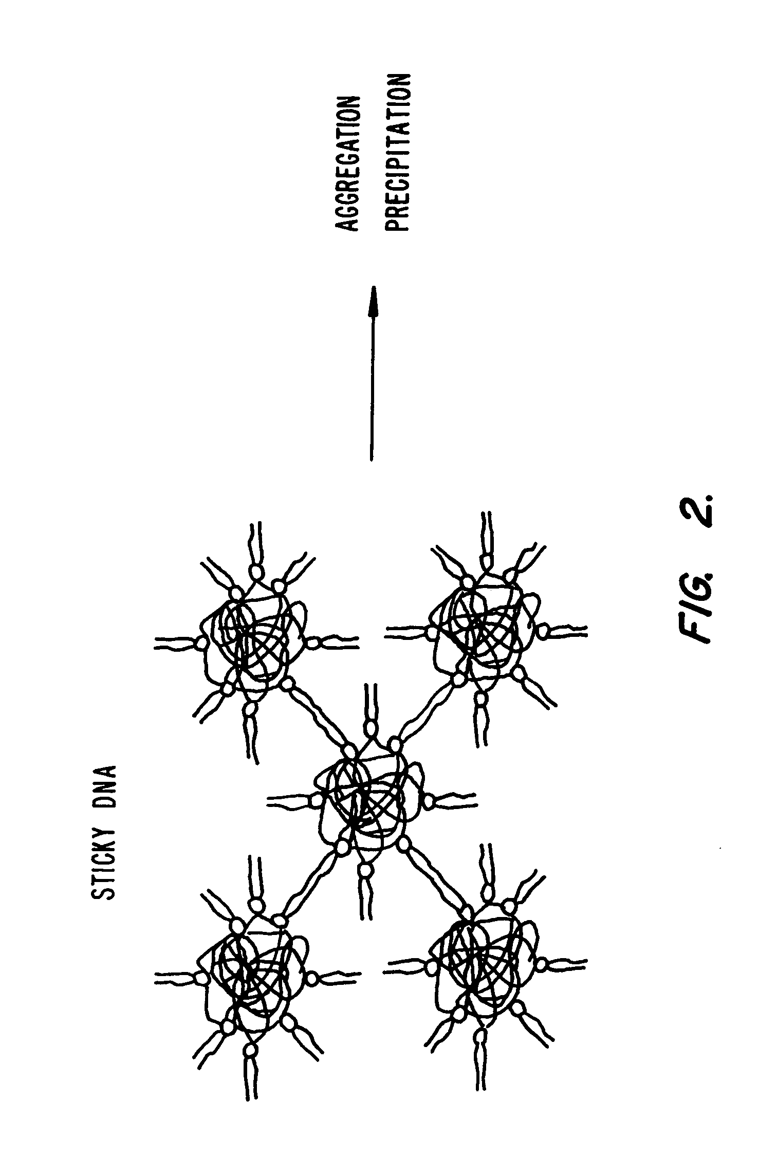
The complexes thus formed have undefined and complicated structures and the lipofection efficiency is severely reduced by the presence of serum.
These complexes are often prepared in the presence of ethanol and are not stable in water.
Additionally, degradation either outside or inside the target cell remains a problem (see, Duzghines, Subcellular Biochemistry 11:195–286 (1985)).

Method used


Image

  • Lipid-nucleic acid particles prepared via a hydrophobic lipid-nucleic acid complex intermediate and use for gene transfer
  • Lipid-nucleic acid particles prepared via a hydrophobic lipid-nucleic acid complex intermediate and use for gene transfer
  • Lipid-nucleic acid particles prepared via a hydrophobic lipid-nucleic acid complex intermediate and use for gene transfer

Examples


example 1

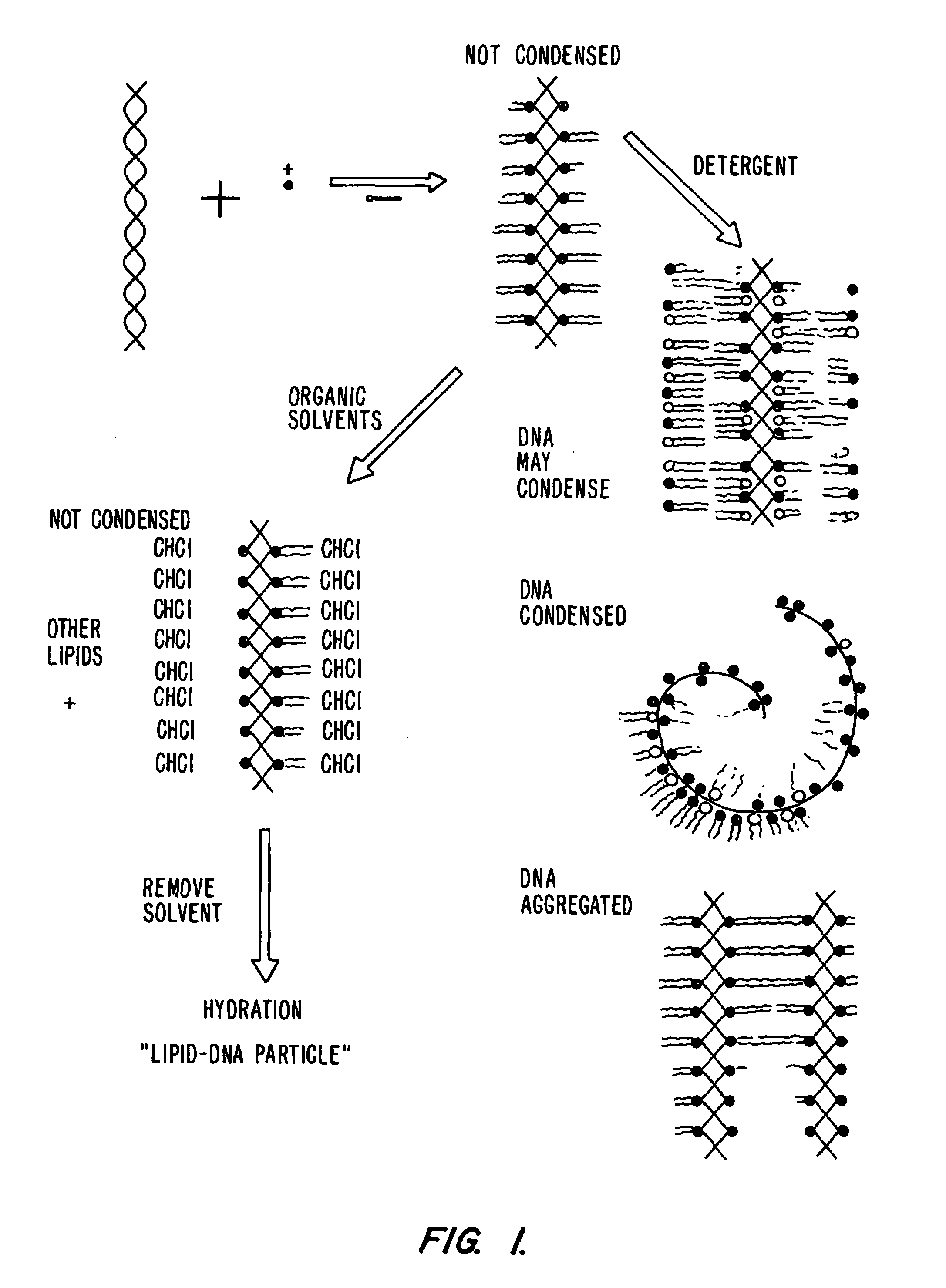
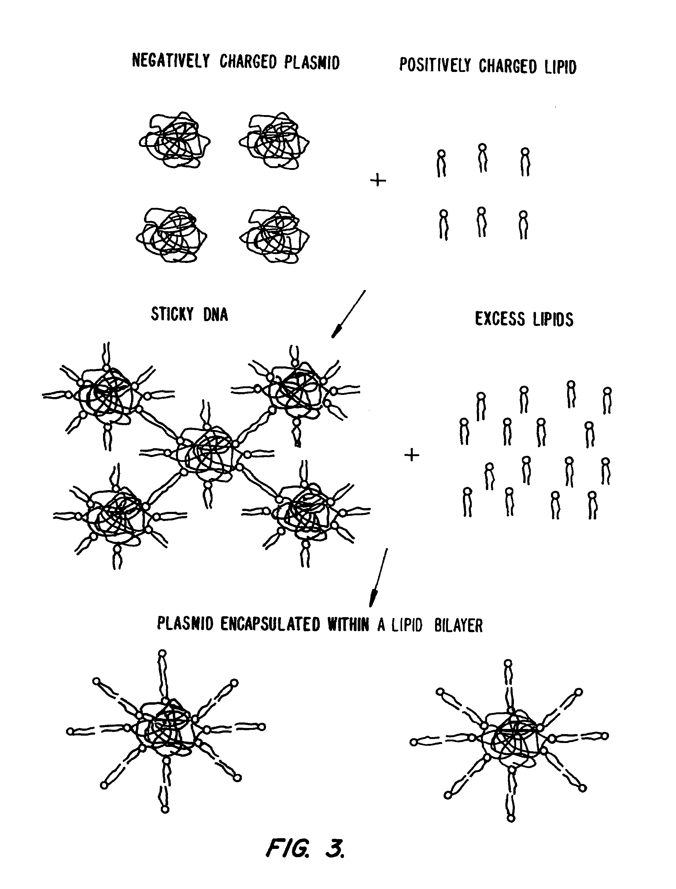
[0251]This example illustrates the encapsulation of a plasmid in a lipid particle using either a reverse-phase method or a detergent dialysis method.

Reverse Phase Method

[0252]pCMV4-CAT plasmid was encapsulated in a lipid particle which was constructed using about 10 mg or 20 mg of lipid. The encapsulation method involved a modification of the classical reverse phase method for entrapment. Generally, 1.050 ml of chloroform:methanol in a 1:2–1 mole % ratio was added to a lipid film containing 2 μl of 14C-cholesteryl hexadecyl ether (6.66 μl / μCi). This was followed by the addition of 220 μl H2O and 33 μl 3H-pCMVCAT plasmid (158,000 dpm / μl; 1.5 mg / ml). This combination provided a clear single phase. The chloroform and most of the methanol were removed under a stream of nitrogen while vortexing. In some cases, the resulting 250 μl suspension of encapsulated plasmid was diluted with 1 ml of H2O and extruded 5 times through one 400 nm filter followed by extrusion 5 times through one 200 nm...

example 2

[0254]This example illustrates the level of plasmid “protection” from the external medium using anion exchange chromatography.

[0255]The extent of encapsulation or protection of the plasmid from the external medium was assessed by anion exchange chromatography as follows: a 50 μl aliquot of each sample was eluted on a DEAE Sepharose CL-6B column and the fractions were assessed for both 3H-plasmid and 14C-lipid by scintillation counting. Any exposed negative charges, such as those present on DNA molecules will bind to the anion exchange column and will not elute with the 14C-lipid. DNA which has its negative charge, “protected” or non-exposed will not bind to the anion exchange resin and will elute with the 14C-lipid. Alternatively, plasmid DNA was measured using the indicator dye, PicoGreen®.

Reverse Phase Method

[0256]FIG. 4 presents the results describing the relationship between the amount of DODAC present in the formulation and the encapsulation efficiency for POPC:DODAC:PEG-Cer-C2...

example 3

[0260]This example illustrates the serum stability achieved using plasmid:lipid particles prepared by the methods of Example 1.

[0261]To establish the serum stability of the plasmid-lipid particles aliquots of the particle mixtures prepared according to both the reverse phase and dialysis method of Example 1 were incubated in 80% mouse serum (Cedar Lane) for 30 min at 37° C. Prior to incubation, the lipid associated plasmid was eluted on a DEAE Sepharose CL-6B column to remove unencapsulated plasmid. Following incubation, an aliquot of the incubation mixture was eluted in HBS on a Sepharose CL-4B column.

[0262]As a control, 1.5 mg of free 3H-pCMVCAT was eluted on a Sepharose CL-4B column in HBS, pH 7.4 (see FIG. 9A). For comparison, 1.5 mg of free 3H—pCMVCAT was incubated in 500 μl of mouse serum at 37° C. for 30 min and eluted in the same manner (FIG. 9B). Note that in FIG. 9A, the free plasmid eluted in the void volume of the column while, in FIG. 9B, the plasmid incubated in serum ...


PUM


Abstract

Novel lipid-nucleic acid particulate complexes which are useful for in vitro or in vivo gene transfer are described. The particles can be formed using either detergent dialysis methods or methods which utilize organic solvents. Upon removal of a solubilizing component (i.e., detergent or an organic solvent) the lipid-nucleic acid complexes form particles wherein the nucleic acid is serum-stable and is protected from degradation. The particles thus formed have access to extravascular sites and target cell populations and are suitable for the therapeutic delivery of nucleic acids.

Description

[0001]This application is a continuation-in-part of U.S. patent application Ser. No. 08 / 485,458, filed Jun. 7, 1995 which issued as U.S. Pat. No. 5,705,385 on Jan. 6, 1998, and of U.S. patent application Ser. No. 08 / 484,282, filed Jun. 7, 1995 which issued as U.S. Pat. No. 5,981,501 on Nov. 9, 1999.FIELD OF THE INVENTION[0002]This invention relates to lipid-nucleic acid particles which are useful for the introduction of nucleic acids into cells, and methods of making and using them. The invention provides a circulation-stable, characterizable delivery vehicle for the introduction of plasmids or antisense compounds into cells. These vehicles are safe, stable, and practical for clinical use.BACKGROUND OF THE INVENTION[0003]Gene transfer into genetically impaired host cells in order to correct the genetic defects has vast potential for successfully treating a variety of thus far hitherto untreatable medical conditions. There are currently six major non-viral methods by which genes are ...

Claims


Application Information

Patent Timeline
Patent Type & Authority Patents(United States)
IPC IPC(8): C12N15/88A01N43/04A61K9/127C07H21/04C12Q1/68
CPCA61K9/1272C12N15/88A61K47/48815Y10S436/829A61K47/6911
Inventor WHEELER, JEFFERY J.BALLY, MARCEL B.ZHANG, YUAN-PENGREIMER, DOROTHY L.HOPE, MICHAELCULLIS, PIETER R.SCHERRER, PETER
Owner TEKMIRA PHARMA CORP
Learn More