
Medical devices

a technology of medical devices and devices, applied in the field of medical devices and systems, can solve the problems of increasing the number of problems associated with known heart valve devices, difficult to implant, and difficult to size properly

Inactive Publication Date: 2008-08-26
L P100
View PDF28 Cites 65 Cited by
  • Summary
  • Abstract
  • Description
  • Claims
  • Application Information

AI Technical Summary

Benefits of technology

[0008]The present invention addresses the abovenamed problems, and, in particular, provides a straightforward, efficacious and economic means of obtaining clinically and physiologically useful in vivo data relating to the condition of a heart valve implant or repair or other indigenous valve treatment, arid of the performance of the heart valve. The in vivo data can be easily obtained on a regular basis over an extended time frame, Further, data can be obtained without requiring the attention of skilled operatives so as to provide the attendant clinical staff with an assessment of any changes, either acute or chronic, with the valve(s) progress since time of implant or treatment.
[0022]At least one sensor may sense blood pressure. In this way, highly relevant clinical pressure data, such as systolic and diastolic pressures, and pressure profiles as a function of time, can be obtained. Advantageously, the medical device comprises at least two spaced apart sensors for sensing blood pressure at different locations in the heart of the patient. In this instance the telemetric communication means may telemetrically transmit data related to the difference in the blood pressures sensed by the at least two sensors. In this way, information on blood flow and blood leakage can be obtained, particularly pressure differences across the valve or valve replacement, giving valuable data concerning valve narrowing / stenosis / incompetence.
[0032]It is highly advantageous that data obtained in vivo from the environs of a heart valve can be conveniently transmitted to a remote device with little or no inconvenience to the patient. Systems of the present invention can be produced economically, thus facilitating mass manufacture arid monitoring of the patient in a wide range of locations such as, for example, a general practitioner's surgery or at the patient's abode. It is a further advantage that the remote device can be a handheld device, thus further facilitating convenient usage. The system does not require the attention of a skilled operative in order to obtain data, and might even be used by the patient himself or herself.
[0033]The remote device may be adapted to provide power remotely to the telemetric communication means. The remote device may be adapted to produce an RF field for this purpose. The remote device may comprise memory for storing data transmitted by the telemetric communication means. The remote device may comprise data analysis means for performing a physiologically or clinically relevant analysis of data transmitted by the telemetric communication means. An example of data analysis is provided by the instance in which two or more sensors sense blood pressure at different locations in the heart of the patient. In this instance, the remote device (or another component in the system) may calculate a quantity related to the difference in the blood pressures sensed by the two or more sensors. This quantity may be integrated with acoustic or other relevant clinical data, thus aiding enhanced clinical evaluation of the performance of the valve or valve replacement.

Problems solved by technology

It is generally recognized that stentless (also known as free sewn or free-stent) tissue valves provide better performance, but suffer from the disadvantages of being rather difficult to implant and difficult to size properly.
However, there are a number of problems associated with known heart valve devices.
At present, this post operative monitoring process may be inconvenient, resource intensive, and expensive.
In particular, monitoring is most efficaciously performed through echocardiography, which requires the provision of expensive ultrasound equipment.
Necessarily, such resources are only maintained at relatively large institutions such as hospitals, and thus require patients to travel (possibly over long distances) to attend a check up, which is inconvenient.
A related problem is that access to echocardiography monitoring is limited in fact, it is the case that implanted heart valve dysfunction associated with abnormal valve action caused by complications such as thrombosis formation or tissue ingrowth tends to develop over a period of several weeks.
However, there is currently no readily available method for performing an in vivo assessment of this type which is not invasive or minimally evasive, or indeed avoids patients and clinicians waiting for appointments for full assessment to be performed.
It is also not easy and somewhat subjective to compare one study with another or to evaluate the gradual changes over a period of time.
It is a disadvantage that the approach is entirely reliant on training the patients to perform an analytical technique and on the ability of the patients to properly perform the technique.

Method used


Image

  • Medical devices
  • Medical devices
  • Medical devices

Examples


Embodiment Construction

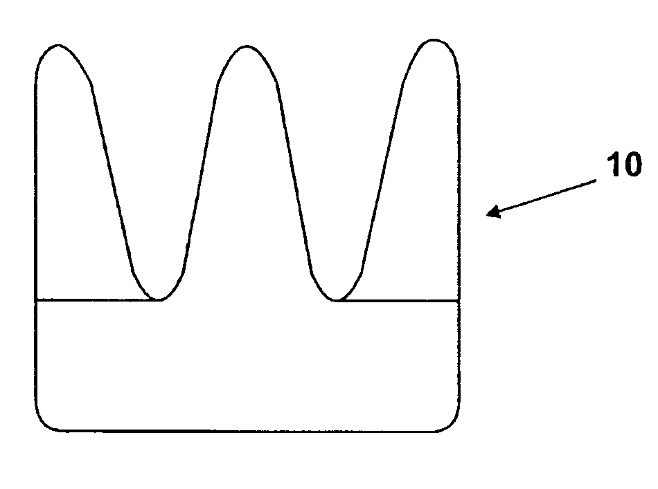
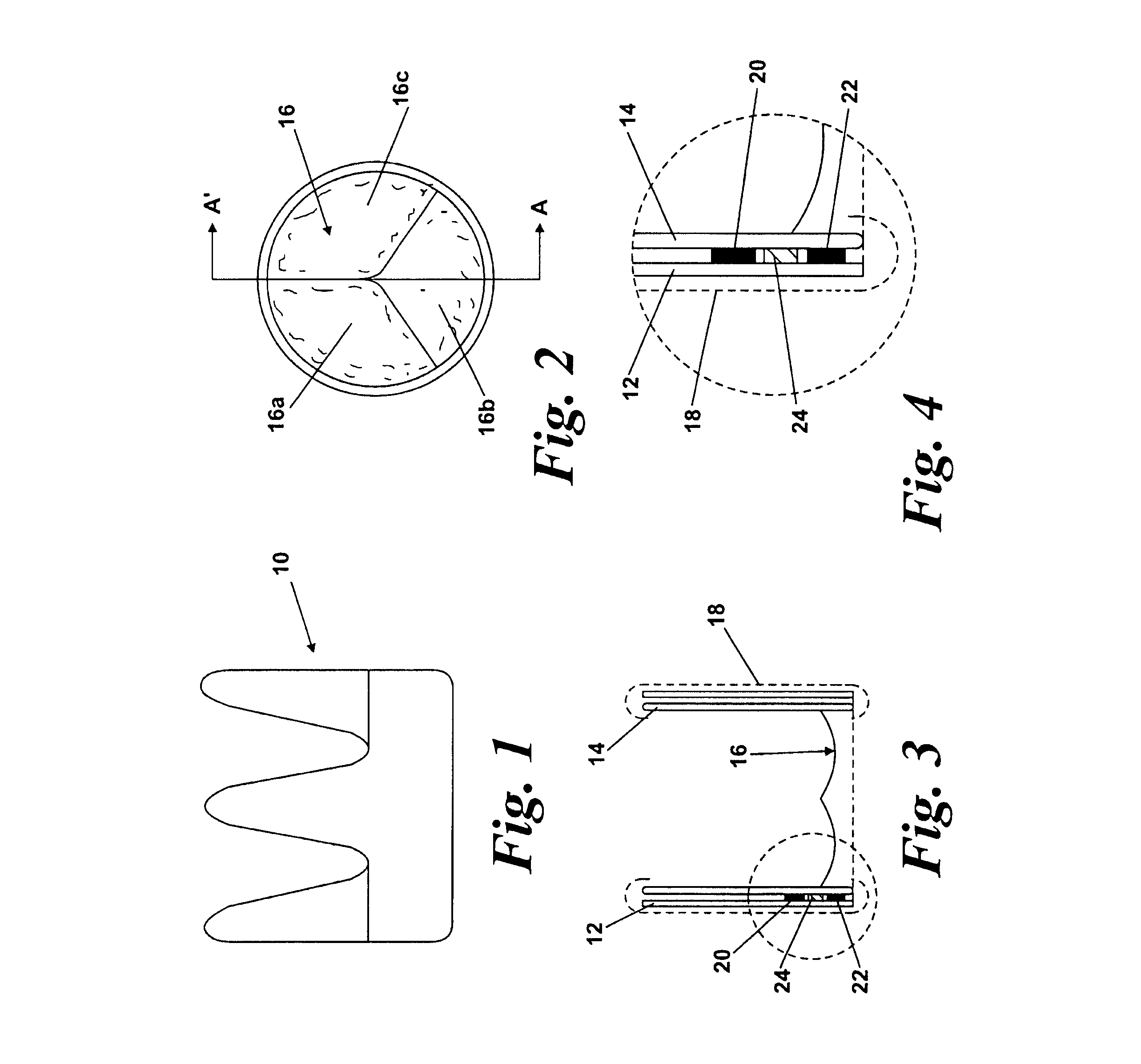
[0048]FIGS. 1-4 show a first embodiment of a heart valve 10 according to the invention. The heart valve 10 is a stented tissue valve comprising a stent 12 which supports a tissue valve wall 14 obtained from a suitable source. Typically, porcine tissue valves are utilized. The tissue valve 10 further comprises a valve 16, the valve 16 being made up of three tissue leaflets 16a, 16b, 16c. Typically, a protective cover 18 is provided around the periphery of the valve wall 14 / stent 12. The cover may be produced from any suitable material: typically, a polymeric sheet material such as Dacron (RIM) is used, although the invention is not limited in this regard. As shown to best effect in FIG. 4, the tissue heart valve 10 further comprises a first sensor 20, a second sensor 22, and telemetric communication means 24, all of which are disposed in the cavity provided between the valve wall 14 and stent 12. This location is extremely convenient, since blood flowing through the tissue heart valv...


PUM


Abstract

There is disclosed a medical device adapted to be implanted in the heart of a patient and operable therein i) as a heart valve; or ii) to assist in the functioning of one of the patient's heart valves; or iii) to monitor the functioning of one of the patient's heart valves. The device includes one or more sensors for sensing a physiologically or clinically relevant parameter of a patient. A telemetric communication device telemetrically transmits data related to a parameter sensed by the sensor to a remote device.

Description

RELATED APPLICATIONS[0001]This application claims the benefit of United Kingdom Patent Application No. 0325679.9, filed on Nov. 4, 2003, which hereby is incorporated by reference in its entirety.[0002]1. Field of the Invention[0003]This invention relates to medical devices and systems that incorporate said medical devices, in particular to heart valve devices.[0004]2. Background of the Invention[0005]Heart valves are well known medical devices which are intended to replicate the function of the valves of the human heart, i.e., to regulate the flow of blood into or out of the chambers of the heart. Known heart valves can be categorized into two main types, namely mechanical heart valves and tissue heart valves. With mechanical valves, the entire valve is constructed from non-living material, which may be a synthetic or man-made material. They may comprise a tilting disc or occluder or incorporate a bileaflet design. Many mechanical valves, both occluders or bileaflet, are made from p...

Claims


Application Information

Patent Timeline
Patent Type & Authority Patents(United States)
IPC IPC(8): A61B5/05A61F2/24A61B5/00A61B5/0215A61B5/07A61B7/00A61F2/02A61F2/06A61F2/07A61F2/86
CPCA61B5/0031A61B5/076A61B5/6862A61B5/6876A61B5/0215A61B5/1473A61F2250/0002A61B2560/0219A61F2/07A61F2/24A61F2/86A61F2002/065A61F2002/075A61B7/005
Inventor TURNER, NICHOLAS MCMAHONPAYNE, PETER ALFREDDAVIES, G. ALBANDAVIES, JULIE A.O'RIORDAN, SIMON JOHN PATRICK
Owner L P100
Learn More