
Apparatus for monitoring a system with time in space and method for diagnosing a condition of a prostate

a technology of time in space and prostate cancer, applied in the field of apparatus for monitoring a system with time in space and diagnosing a prostate condition, can solve the problems of prostate cancer being a major socioeconomic problem, difficult to achieve high spatial resolution and maintain high temporal resolution, etc., and achieve high angiogenicity, reduce fluid content, and high cellularity

Inactive Publication Date: 2007-07-17
YEDA RES & DEV CO LTD
View PDF2 Cites 49 Cited by
  • Summary
  • Abstract
  • Description
  • Claims
  • Application Information

AI Technical Summary

Benefits of technology

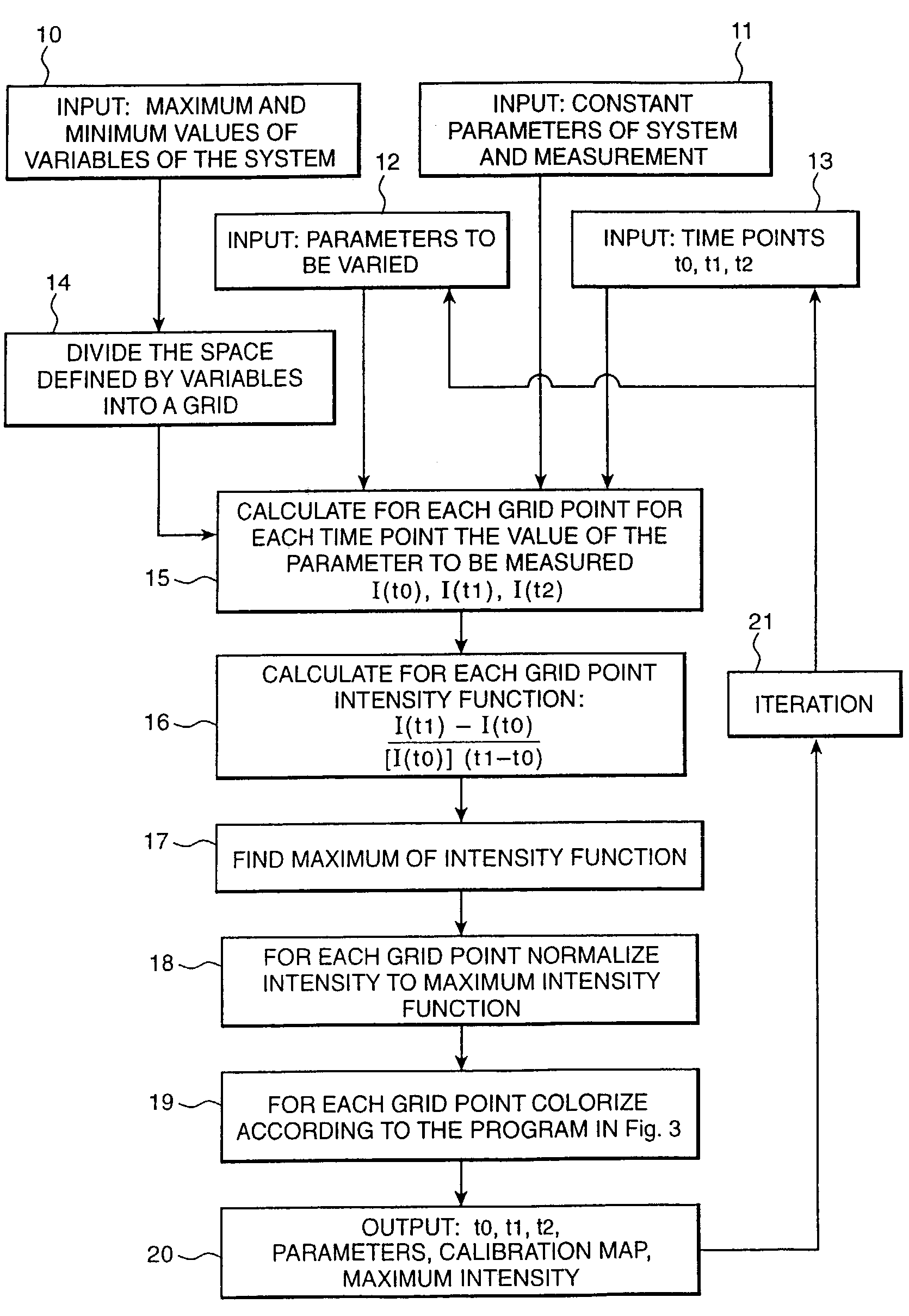
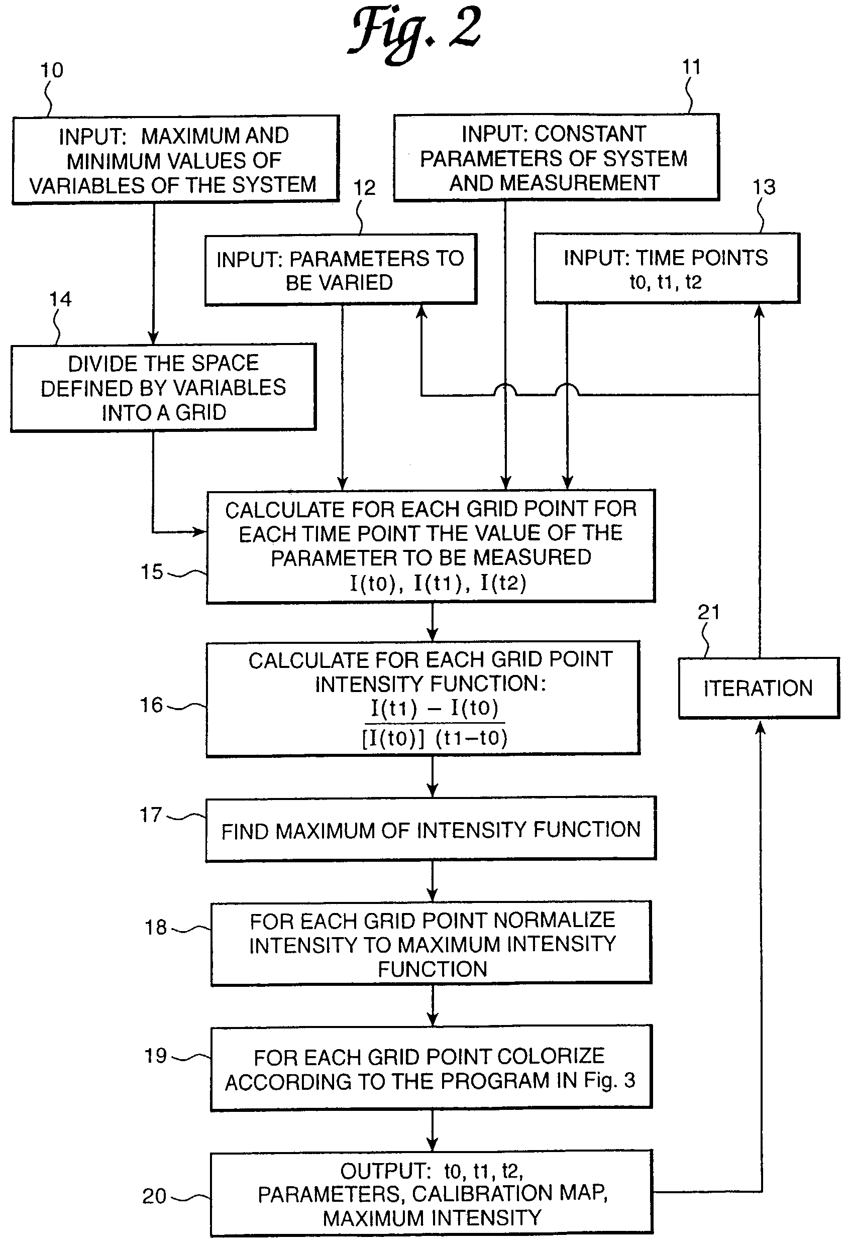
[0025]The principles of the 3TP method are general and can be applied to monitor prostate cancer patients as well as other diseases that demonstrate contrast changes following contrast agent administration. Blood vessel density in prostate cancer is an independent indicator of pathological state and prognosis. With the addition of vessel permeability by the 3TP method, which reflects high angiogenic activity, this parameter may be even more reliable as a prognostic factor. The color distribution in the 3TP images also enables characterizing tumor cellularity (the density of the cells in the tumor) and hence the differentiation of the lesion with high cellularity. The reduced fluid content of well to moderately well differentiated tumors can be further used to improve the specificity of tumor staging.
[0026]By enhancing the in-plane resolution and performing a pixel-by-pixel model-based analysis, the 3TP method can be optimized to improve tissue characterization and staging accuracy of prostate MRI. It is expected that tumor volume will be also assessed more accurately. A high percentage of red bright areas in the 3TP images of cancers may indicate a worse prognosis than for those with a low percent. The exact correlation between the color hue and color intensity for assessment of prognosis can be obtained by a clinical trial. In summary, contrast-enhanced MRI with the 3TP method for the dynamic data analysis can provide useful staging information. The technique may be performed together with conventional T2-weighted images within a clinically acceptable examination time.

Problems solved by technology

In a standard clinical MRI of the breast, it is difficult to achieve high spatial resolution and also maintain high temporal resolution.
As a consequence, in standard MRI machines the output results are sometimes inconclusive.
This high prevalence, and a rising age-adjusted mortality rate, makes prostate cancer a major socioeconomic problem.
Even with 13 cores, however, it is still possible to completely miss areas of carcinoma, and hence, understage the disease.
However, this distinction is often lost in the prostate and depends on the location of the pathologic changes within the prostate.
Due to similarities between tumor and central gland tissues, poor discrimination results, particularly in the presence of coexisting benign disease.
The use of contrast-enhanced MRI to diagnose prostate diseases has so far been limited.
It was indicated that the low spatial resolution in these studies is a serious limiting factor, especially when dealing with heterogeneous lesions.
For these patients, the possible risk and side-effects of surgery and treatment may outweigh the possible benefits of treatment.
The decision as to who will benefit from a prostatectomy is a major issue.
Clearly, some of these men die before their prostate carcinoma causes significant problems and such tumors have been described as “insignificant.” Others undergo a radical prostatectomy only to discover that their tumors have grown beyond the prostate, and 50% of these men will suffer a recurrence.
Currently, however, there is no precise clinical method for the reliable assessment of local extent of prostate cancer.
This is a major drawback in the treatment of localized disease.

Method used


Image

  • Apparatus for monitoring a system with time in space and method for diagnosing a condition of a prostate
  • Apparatus for monitoring a system with time in space and method for diagnosing a condition of a prostate
  • Apparatus for monitoring a system with time in space and method for diagnosing a condition of a prostate

Examples


examples

Parametric MRI of Tumor Perfusion; from Cellular Studies to Patient's Diagnosis

[0090]The growth of solid tumors relies on their perfusion through their vasculature and is determined by the volume fraction of the capillaries as well as the capillary flow and permeability. Magnetic resonance imaging can be applied in vivo for characterizing tumor perfusion and thereby provide a unique tool for monitoring and understanding the activity of angiogenic growth factors and suppressors.

[0091]Dynamic high resolution 1H imaging and 2H imaging methods were developed and applied to investigate orthotopically implanted human prostate cancer in mice. Processing algorithms based on mathematical models of the dynamic behavior were developed and applied at pixel resolution. The final analyses were presented as images of physiologic parameters (parametric images) that characterized uniquely tumor perfusion. The parametric images obtained by both methods revealed the high heterogeneity of cancer perfus...


PUM


Abstract

Apparatus and method for monitoring a prostate in a human body in which a fluid flows and which is characterized by a change in the prostate with time in space for the purpose of diagnosing a prostate condition, particularly cancer. A preselected place in the prostate is monitored to collect data at two or more time points correlated to a prostate event. The data is indicative of a prostate parameter that varies with time as a function of at least two variables related to prostate wash-in and wash-out behavior. A calibration map is made on a calculated basis with each pixel or voxel representative of a color hue indicative of wash-out behavior and a color intensity indicative of wash-in behavior. When a satisfactory map is obtained, the collected data is processed on the basis of the map to obtain an image of the preselected place with each spatial unit thereof correlated with a color hue and a color intensity. Software and a data processing system are provided to develop the calibration map. The calibration map and image of the preselected place are also novel implementations.

Description

[0001]This application is a Continuation-in-Part of U.S. application Ser. No. 09 / 993,190 filed Nov. 16, 2001, now U.S. Pat. No. 6,611,778 which is a divisional application of Ser. No. 09 / 101,708 filed Sep. 16, 1998, now U.S. Pat. No. 6,353,803 granted Mar. 5, 2002, which was a National Stage filing of PCT Application Number PCT / US97 / 00801 filed Jan. 21, 1997.FIELD OF THE INVENTION[0002]The present invention relates to apparatus for monitoring a system with time in space and method for diagnosing a condition of a prostate, especially cancer, and more particularly relates to an apparatus such as a unique MRI machine for diagnosing a condition of a prostate, especially cancer.BACKGROUND OF THE INVENTION[0003]Presently apparatuses are known for monitoring testing or measuring a system in which a fluid that is flowing or substances in the fluid will dissipate in part as it traverses the system or will require regeneration. For example, MRI machines are used today to create images with or...

Claims


Application Information

Patent Timeline
IPC IPC(8): G06K9/00G01F1/12G01G23/01G01N15/08G06T15/70
CPCG06T7/0012G06T2207/10088G06T2207/30081G06T2207/30104
Inventor DEGANI, HADASSAFURMAN-HARAN, EDNA
Owner YEDA RES & DEV CO LTD
Learn More