
Method for diagnosing preeclampsia by detecting hCRH mRNA

a preeclampsia and hcrh technology, applied in the field of preeclampsia diagnosis by detecting hcrh mrna, can solve the problems of limited application of this technology, increased complexity of fetal dna-based analysis, and invasive conventional methods, so as to increase the risk of developing, increase the amount of mrna, and increase the mrna level

Active Publication Date: 2007-06-26
THE CHINESE UNIVERSITY OF HONG KONG
View PDF8 Cites 32 Cited by
  • Summary
  • Abstract
  • Description
  • Claims
  • Application Information

AI Technical Summary

Benefits of technology

[0143]SUPPLEMENTARY TABLE AMicroarray detection of the 50 most highly expressed genes in CVS tissuesTranscriptProbe Set IDGenBank acc.Signals (median)*chorionic gonadotropin, beta polypeptide205387_s_atNM_000737.131857.3placental lactogen (v.4)208356_x_atNM_022642.128651.4growth hormone 1 (v.5)208068_x_atNM_022562.128500.7growth hormone 1 (v.4)208069_x_atNM_022561.127624.7chorionic somatomammotropin hormone 2 (v.1)203807_x_atNM_020991.227513.2placental lactogen (clone MGC:14518)211739_x_atBC005921.127185.9chorionic somatomammotropin hormone-like 1 (v.2)208294_x_atNM_022578.127104.4*placental lactogen (v.1)202493_x_atNM_001317.226503.0chorionic somatomammotropin hormone 2 (v.3)208342_x_atNM_022645.125820.6placental lactogen (v.3)208357_x_atNM_022641.125777.3chorionic somatomammotropin hormone 2 (v.4)208341_x_atNM_022646.125603.1pregnancy specific beta-1-glycoprotein 1208257_x_atNM_006905.125453.4growth hormone 1 (v.2)206885_x_atNM_022559.124834.8chorionic somatomammotropin hormone-like 1 (v.1)207285_x_atNM_001318.224796.9chorionic somatomammotropin hormone-like 1 (v.5)208293_x_atNM_022581.124709.4pregnancy specific beta-1-glycoprotein 6209738_x_atM31125.124370.6pregnancy specific beta-1-glycoprotein 3203399_x_atNM_021016.124262.4chorionic somatomammotropin hormone-like 1 (v.4)208295_x_atNM_022580.124190.7*tissue factor pathway inhibitor 2209278_s_atL27624.124179.9glycoprotein hormones, alpha polypeptide204637_atNM_000735.223720.4growth hormone 1 (v.3)206886_x_atNM_022560.123475.7pregnancy specific beta-1-glycoprotein 3215821_x_atAB019570.123095.5pregnancy specific beta-1-glycoprotein 3211741_x_atBC005924.123025.1growth hormone variant mRNA211151_x_atAF185611.122792.2prostate differentiation factor221577_x_atAF003934.122685.4growth hormone 1 (v.1)205840_x_atNM_000515.222446.6chorionic somatomammotropin hormone-like 1 (v.3)205958_x_atNM_022579.122097.6pregnancy specific beta-1-glycoprotein 4208191_x_atNM_002780.120730.7pregnancy specific beta-1-glycoprotein 2208134_x_atNM_031246.120233.5pregnancy specific beta-1-glycoprotein 9209594_x_atM34421.119939.4*KiSS-1 metastasis-suppressor205563_atNM_002256.119755.8placental lactogen (v.2)206475_x_atNM_022640.119367.1pregnancy specific beta-1-glycoprotein 5204830_x_atNM_002781.119182.6growth hormone 2211508_s_atAF006060.118019.7S100 calcium binding protein P204351_atNM_005980.117970.2pregnancy specific beta-1-glycoprotein 6208106_x_atNM_002782.317566.8chorionic somatomammotropin hormone 2 (v.2)207770_x_atNM_022644.117244.2CD63 antigen (melanoma 1 antigen)200663_atNM_001780.117061.2delta-like 1 homolog (Drosophila)209560_s_atU15979.116415.0fibronectin 1210495_x_atAF130095.116046.9Homo sapiens cDNA: FLJ22066 fis, clone HEP10611202409_atX0786816024.8fibronectin 1216442_x_atAK026737.115834.1ribosomal protein L31200963_x_atNM_000993.115252.6a disintegrin and metalloproteinase domain 12 (v.2)204943_atNM_021641.115007.3collagen, type III, alpha 1 (clone HEMBA1001071)215076_s_atAU14416714999.1pregnancy specific beta-1-glycoprotein 9207733_x_atNM_002784.114819.7secreted phosphoprotein 1209875_s_atM83248.114665.3Epstein-Barr virus induced gene 3219424_atNM_005755.114617.0collagen, type III, alpha 1201852_x_atAI81375814494.7fibronectin 1211719_x_atBC005858.114157.1v, transcripts variant; * transcripts selected for QRT-PCR study*cortocotropin releasing hormone is located at position 156 on this list*placenta-specific 1 is located at position 412 on this list

Problems solved by technology

These conventional methods are, however, invasive and present an appreciable risk to both the mother and the fetus despite most careful handling (Tabor et al., Lancet 1:1287–1293, 1986).
This approach has limited the application of this technology to the 50% of pregnant women who are carrying male fetuses.
Further, the use of other genetic polymorphisms has also increased the complexity of fetal DNA-based analyses.

Method used


Image

  • Method for diagnosing preeclampsia by detecting hCRH mRNA
  • Method for diagnosing preeclampsia by detecting hCRH mRNA
  • Method for diagnosing preeclampsia by detecting hCRH mRNA

Examples


example 1

Elevated hCRH mRNA Level in Preeclamptic Women

A. Methods

[0064]Subjects

[0065]Peripheral blood samples were collected with informed consent and Research Ethics Committee approval from pregnant women, who attended the Department of Obstetrics and Gynecology at the Prince of Wales Hospital, Hong Kong.

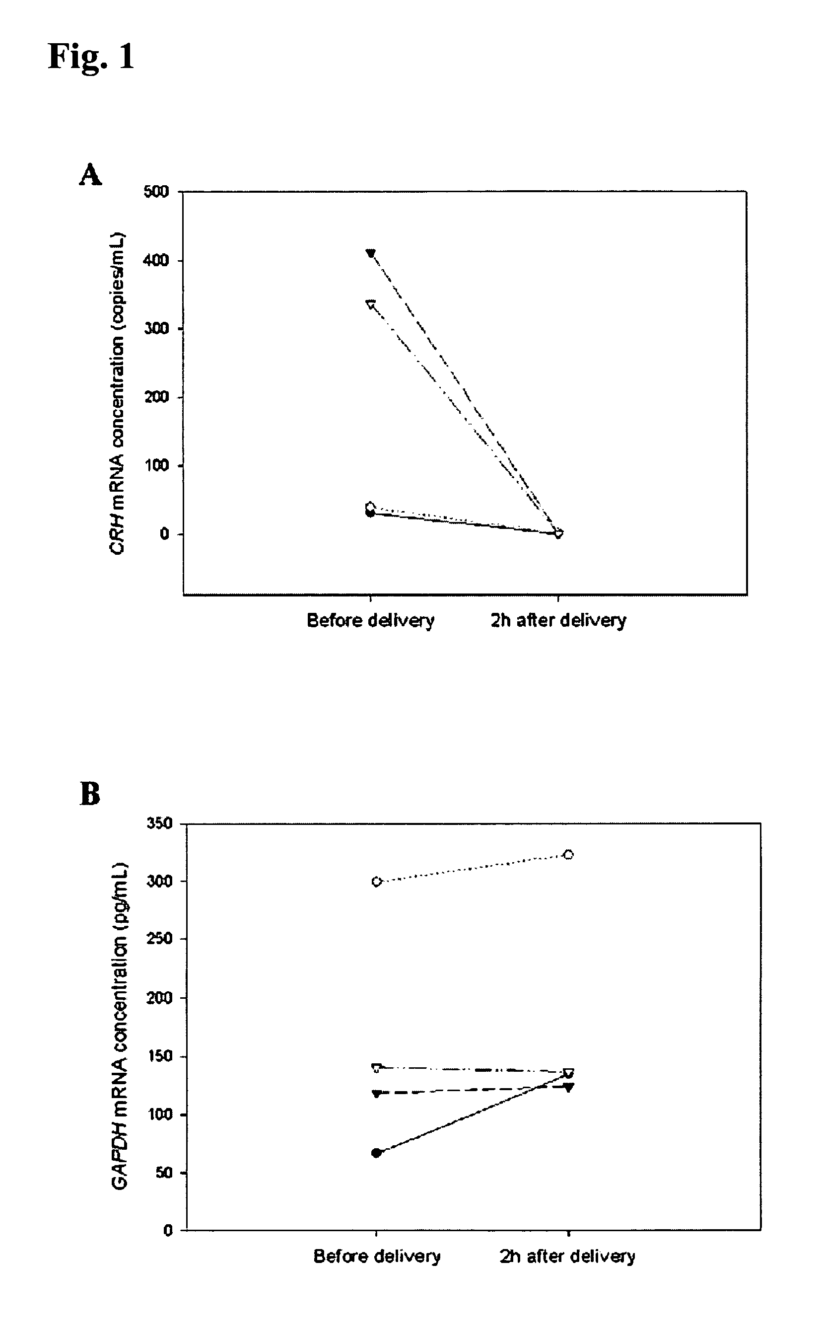
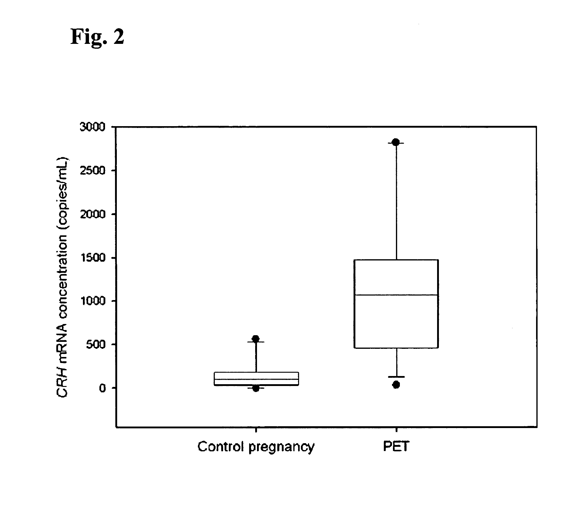
[0066]In the first part of this study, blood samples were obtained from 10 healthy pregnant women during the third trimester of gestation. In the second part of the project, 4 pregnant women with uncomplicated pregnancy were recruited just prior to elective cesarean section. Peripheral blood samples were taken from these subjects just prior to delivery and at 2 hours post-delivery. In the third part of the study, two patient groups were studied: (a) 12 preeclamptic women and (b) 10 control pregnancies. The median gestational ages of the preeclamptic and control groups were 37 weeks and 38 weeks, respectively. Preeclampsia was defined on the basis of a sustained increase in diastolic blood p...

example 2

Detection of hPL and hCG-β mRNA in the Plasma of Pregnant Women

A. Methods

[0086]Subjects

[0087]15-ml blood samples were collected with informed consent and Research Ethics Committee approval from healthy women with singleton uncomplicated pregnancies, who attended the Department of Obstetrics and Gynecology at the Prince of Wales Hospital, Hong Kong.

[0088]Processing of Blood Samples

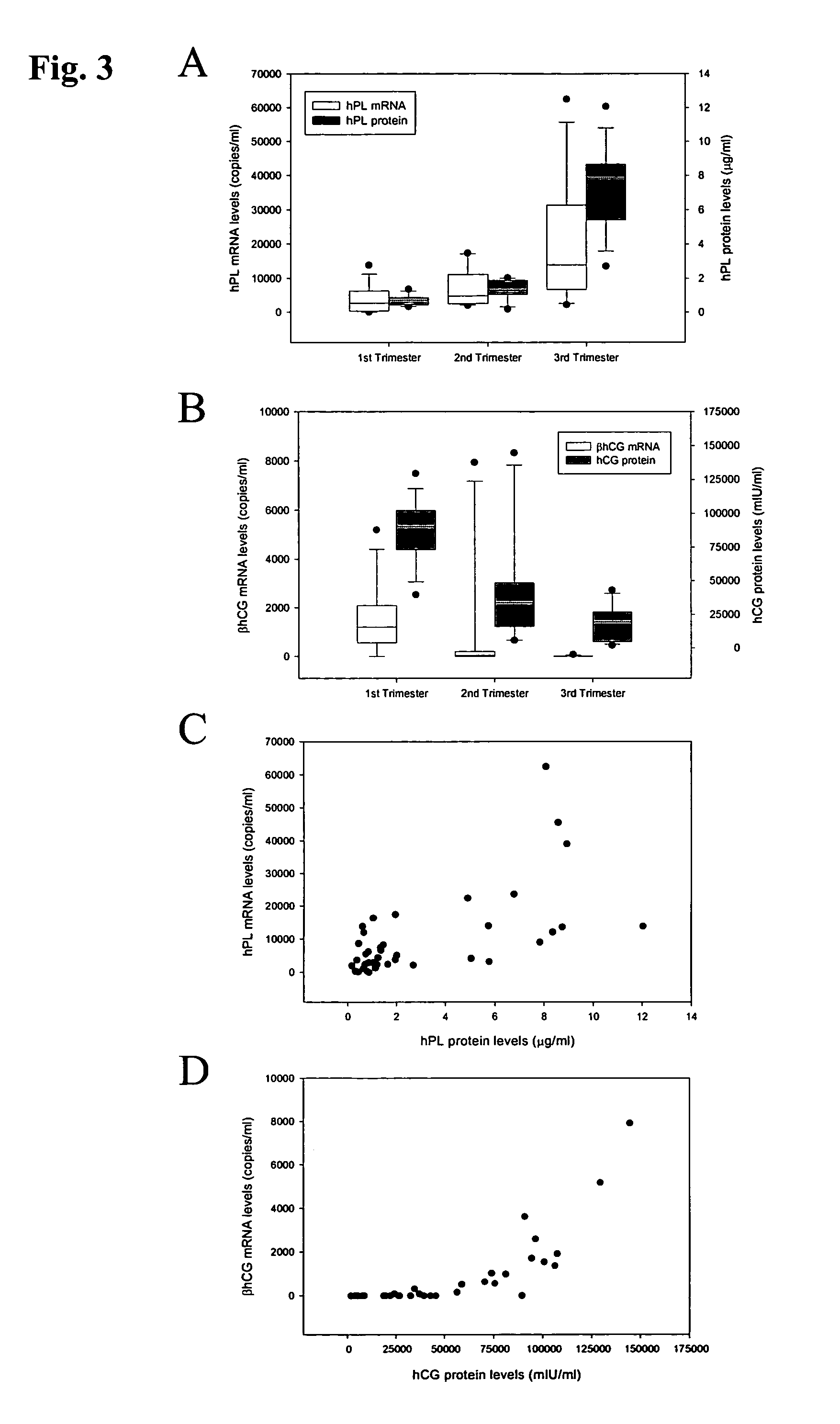
[0089]The blood samples were collected in EDTA-containing and plain tubes, and centrifuged at 1600×g for 10 min at 4° C. Plasma and serum were then carefully transferred into plain polypropylene tubes. The serum samples were stored at −20° C. for immunoassays for the hPL and hCG-β proteins. The plasma samples were re-centriftiged at 16000×g for 10 min at 4° C., and the supernatants were collected into fresh polypropylene tubes. All placental tissue samples were immediately stored in an RNA Later stabilizing solution (Ambion, Austin, Tex.) and kept at −80° C. until RNA extraction. For the filtration study, p...

example 3

Elevation in Maternal Plasma GAPDH mRNA in Preeclamptic Women

[0106]Pregnant women attending the Department of Obstetrics and Gynecology at the Prince of Wales Hospital were recruited with informed consent. Preeclampsia was diagnosed using the criteria as indicated in Example 1.

A. Methods

[0107]Maternal blood samples were taken into heparinized tubes and processed as indicated in Example 1. Plasma RNA was extracted and GAPDH mRNA level was quantified as indicated in Example 1.

B. Results

[0108]Elevation in maternal plasma GAPDH mRNA concentrations was observed in the preeclamptic group, when compared with the control group (FIG. 5). The median maternal plasma GAPDH mRNA concentrations in control and preeclamptic subjects were 70 pg / ml and 281 pg / ml, respectively. The difference is statistically significant (Mann-Whitney test, p<0.005).

C. Conclusion

[0109]Maternal plasma GAPDH mRNA is a new noninvasive marker for preeclampsia.


PUM

PropertyMeasurementUnit
temperatureaaaaaaaaaa
diastolic blood pressureaaaaaaaaaa

Abstract

Methods and kits are provided for diagnosing, monitoring, or predicting the conditions of pre-eclaimpsia, fetal chromosomal aneuploidy, and pre-term labor in a pregnant woman, as well as for detecting pregnancy in a woman, by quantitatively measuring in the maternal blood the amount of one or more mRNA species encoding human chorionic gonadotropin β subunit (hCG-β), human placental lactogen (hPL), human corticotropin releasing hormone (hCRH), KiSS-1 metastasis-suppressor (KISS1), tissue factor pathway inhibitor 2 (TPFI2), placenta-specific 1 (PLAC1), or glyceraldehyde-3-phosphate dehydrogenase (GAPDH), and comparing the amount of the mRNA species with a standard control.

Description

RELATED APPLICATIONS[0001]This application claims priority to U.S. Provisional Application Ser. No. 60,440,906, filed Jan. 17, 2003, the content of which is incorporated herein by reference in the entirety.BACKGROUND OF THE INVENTION[0002]Prenatal diagnosis has been routinely conducted using cells isolated from the fetus through procedures such as chorionic villus sampling (CVS) or amniocentesis. These conventional methods are, however, invasive and present an appreciable risk to both the mother and the fetus despite most careful handling (Tabor et al., Lancet 1:1287–1293, 1986).[0003]Alternatives to these invasive approaches have been developed for prenatal screening, e.g., to detecting fetal abnormalities, following the discoveries that several types of fetal cells can be found in maternal circulation (Johansen et al., Prenat. Diagn. 15:921–931, 1995) and more importantly, circulating cell-free fetal DNA can be detected in maternal plasma and serum (Lo et al., Lancet 350:485–487, ...

Claims


Application Information

Patent Timeline
Patent Type & Authority Patents(United States)
IPC IPC(8): C12Q1/68C07H21/02C07H21/04C12P19/34
CPCC12Q1/6883C12Q2600/158C12Q2600/156
Inventor LO, YUK-MING DENNISNG, KAI ONTSUI, BO YINCHIU, WAI KWUN ROSSA
Owner THE CHINESE UNIVERSITY OF HONG KONG
Learn More