
System and method for identifying optimized blood signal in medical images to eliminate flow artifacts

a technology of optimized blood signal and flow artifact, applied in image enhancement, instruments, applications, etc., can solve the problems of frequent corruption of arteries, and achieve the effect of eliminating flow artifacts and improving accuracy

Active Publication Date: 2007-06-19
VIRTUALSCOPICS
View PDF13 Cites 4 Cited by
  • Summary
  • Abstract
  • Description
  • Claims
  • Application Information

AI Technical Summary

Benefits of technology

[0007]It is an object of the invention to allow the calculation of blood uptake curves with improved accuracy. It is another object of the invention to allow such calculation such that flow artifacts are eliminated.
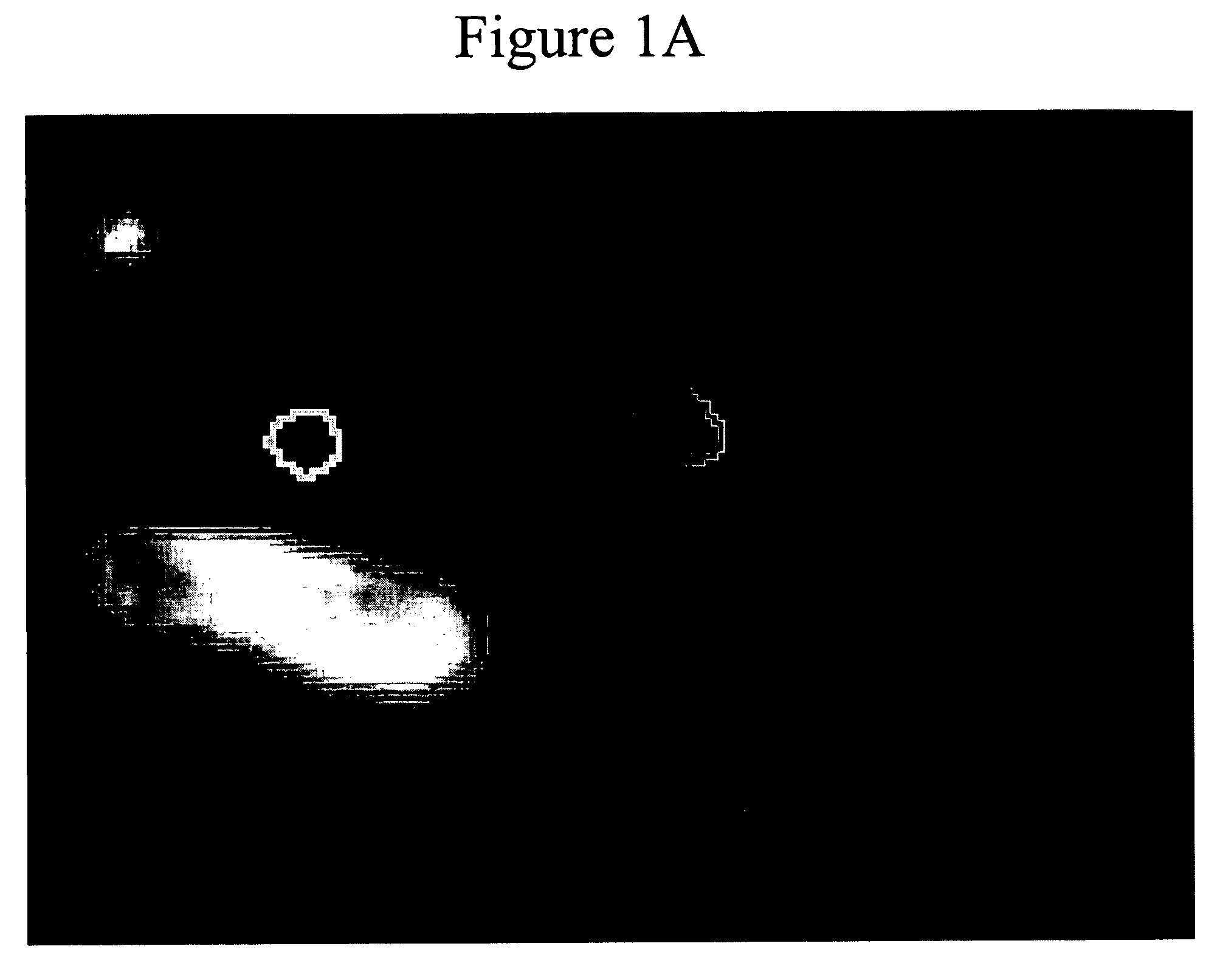
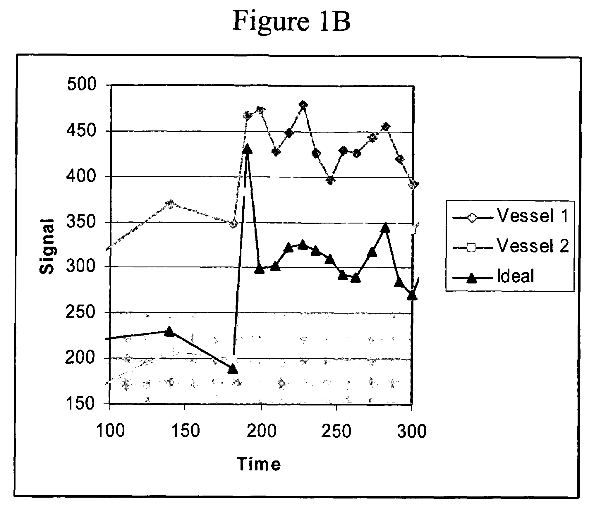
[0008]To achieve the above and other objects, the present invention is directed to a system and method for the identification of an optimized plasma signal which is intended to eliminate this source of measurement variability and thereby increase the sensitivity to change of perfusion parameter measurements. The uptake curve according to the present invention shows a greater enhancement peak than that of either vessel, a smoother plateau, and a more characteristic shape.
[0010]Manual plasma identification for perfusion parameter calculation is currently standard practice for both clinical and experimental purposes. Increased accuracy and sensitivity to change can be achieved by making use of an automated method for plasma identification, as achieved by the present invention.

Problems solved by technology

One of the primary challenges in estimating perfusion parameters is identifying an accurate plasma uptake curve.
However, the MR signal in arteries is frequently corrupted by flow artifacts, with the result that regions of interest at different points in the same artery or in other nearby vessels can provide grossly different uptake curves.

Method used


Image

  • System and method for identifying optimized blood signal in medical images to eliminate flow artifacts
  • System and method for identifying optimized blood signal in medical images to eliminate flow artifacts
  • System and method for identifying optimized blood signal in medical images to eliminate flow artifacts

Examples


Embodiment Construction

[0023]A preferred embodiment of the present invention, as well as experimental results, will now be set forth in detail with reference to the drawings.

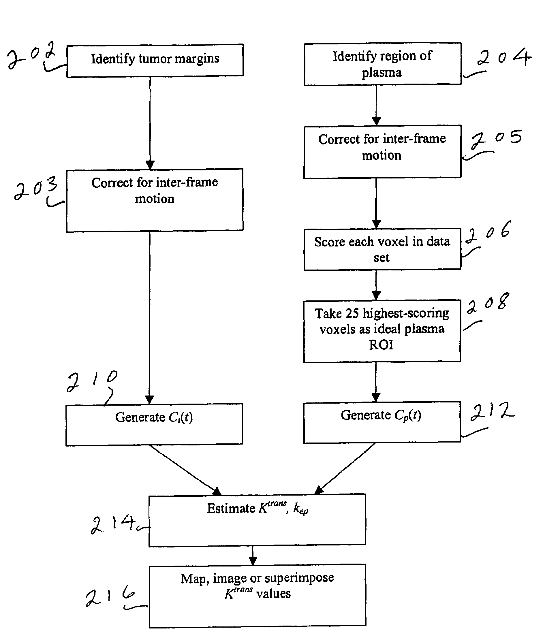
[0024]The process according to the preferred embodiment will be disclosed with reference to the flow chart of FIG. 2. Tumor margins are identified in step 202 using Geometrically Constrained Region Growth (GEORG). This technique requires a user to place a seed or string of seeds within each desired structure throughout the volume using one or more mouse clicks. The seed regions then expand into neighboring voxels provided that two constraints are satisfied: the grayscale value of the neighboring voxel must have a high probability of falling within the statistical distribution defined by all currently included voxels, and inclusion of the neighboring voxel must not cause the shape of the included region to deviate excessively from the a priori regional shape model. Once initiated, the expansion process continues until a stable boundary...


PUM


Abstract

In a sequence of medical image data showing tumors and blood vessels, a plasma signal is optimized to avoid flow artifacts by receiving a user input of a blood region and using the user input to seed an automated search. Each voxel is scored by time point of maximum intake, slope at maximum intake, peak value and conformance to a gamma variate curve, and the voxels with the highest scores are included in the ideal plasma region of interest. Uptake curves for both tumors and plasma are determined and used to estimate a volume transfer constant.

Description

FIELD OF THE INVENTION[0001]The present invention is directed to the identification of blood signals in medical images, such as magnetic resonance (MR) images, and more particularly to such identification which overcomes the corruption of such signals by arterial flow artifacts.DESCRIPTION OF RELATED ART[0002]Dynamic contrast enhanced MRI (dceMRI) has demonstrated considerable utility in both diagnosing and evaluating the progression and response to treatment of malignant tumors. dceMRI involves the periodic acquisition of T1-weighted images before, during and after injection of a gadolinium labeled tracer such as gadopentetate dimeglumine. The change over time in signal intensity in a voxel or region of interest in this time series can then be related to tracer concentration. By making use of a two-compartment model, with one compartment representing blood plasma and the other abnormal extra-vascular extra-cellular space (EES), the observed uptake curves in tissue and plasma can be...

Claims


Application Information

Patent Timeline
Patent Type & Authority Patents(United States)
IPC IPC(8): G06K9/00A61B5/05A61B5/055A61B6/03G01R33/561G01R33/563
CPCG06T7/0083G01R33/56366G01R33/5601G01R33/56308A61B6/507G06T2207/30101G06T7/12
Inventor ASHTON, EDWARD
Owner VIRTUALSCOPICS
Learn More