
Method for diagnosing a transmissible spongiform subacute encephalyopathy caused by an unconventional transmissible agent strain in a biological sample

Inactive Publication Date: 2006-08-29
COMMISSARIAT A LENERGIE ATOMIQUE ET AUX ENERGIES ALTERNATIVES
View PDF13 Cites 19 Cited by
  • Summary
  • Abstract
  • Description
  • Claims
  • Application Information

AI Technical Summary

Benefits of technology

[0060]in the context of method B, the aim of controlling the degradation by the protease will be to degrade the majority, and optionally all, of the octapeptide motif repeats for the PrPs-res corresponding to the strain(s) of interest, whatever the species in which it (they) is (are) expressed, under conditions in which all or some of the octapeptide motif repeats are conserved for the PrPs-res corresponding to all the other TSSE strains. In this case, the advantage of the invention is to allow differential diagnosis of the strain of interest relative to the other UTA strains.
[0062]Surprisingly, under the conditions according to step (1) or step (a), the PK does not lead to the cleavage of all or some of the octapeptide motif repeats of the PrPs-res associated with all UTA strains or prions, whereas the conditions of step (b) lead to the cleavage of the octapeptide motif repeats of the PrP-res associated with the strain of interest, while all the PrPs-res associated with the other UTA strains conserve all or some of said octapeptide motif repeats.
[0063]Also surprisingly, such a test, in particular when the ligand is an antibody, has the following advantages:
[0064]great sensitivity of detection since the antibodies directed against these octapeptide motif repeats have a much greater affinity than the antibodies directed against other regions of the PrP-res 27-30 and are more resistant to the buffers used; specifically, the recognition of a repeat motif promotes interactions of high affinity and provides the possibility of attaching several antibody molecules to a single PrP molecule;
[0065]the possibility of differential diagnosis of the UTA strains or prions since it is possible, depending on the conditions used, to obtain or not to obtain digestion of these motifs; it is in particular possible to eliminate them for all the diseases linked to the strain of interest, the agent for BSE for example, whereas it is possible to conserve it for the other “prion” diseases. As a result of this, the methods according to the invention provide a simple test for the differential diagnosis of BSE and / or of another strain of interest, relative to the other strains, by using two different conditions (step (1) or (a) and step (b)), the conditions of step (1) or (a) (conserving the octapeptide motif repeats of the PrPs-res associated with all UTA strains) allowing the detection of all the strains, and the conditions of step (b) (eliminating these motifs only in the PrP-res associated with a strain of interest) revealing only the other strains.
[0066]Such tests therefore in particular find an application in the investigation of the contamination of sheep with the BSE strain, which is not usually possible to distinguish from the conventional strains of scrapie.

Problems solved by technology

These diseases are always fatal and, at the current time, there is no effective treatment.
In vitro, it is toxic for cultures of neurons.
In general, these various tests have the drawback of lacking sensitivity and thus of causing false-negatives.
The method described in WO 00 / 29850, Wallac Oy et al., has the major drawback of lacking specificity, due, in particular, to incomplete elimination of PrP-sen under the recommended treatment conditions, while the method described in WO 00 / 22438, The Regents of the University of California, lacks sensitivity due to the use of the detecting antibody 3F4 (WO 00 / 22438), which binds to only one motif on the protein.

Method used


Image

  • Method for diagnosing a transmissible spongiform subacute encephalyopathy caused by an unconventional transmissible agent strain in a biological sample
  • Method for diagnosing a transmissible spongiform subacute encephalyopathy caused by an unconventional transmissible agent strain in a biological sample
  • Method for diagnosing a transmissible spongiform subacute encephalyopathy caused by an unconventional transmissible agent strain in a biological sample

Examples


example 1

Production and Characterization of Monoclonal Antibodies Specific for the Octapeptide Motif Repeat

Synthesis and Labeling of the Peptide

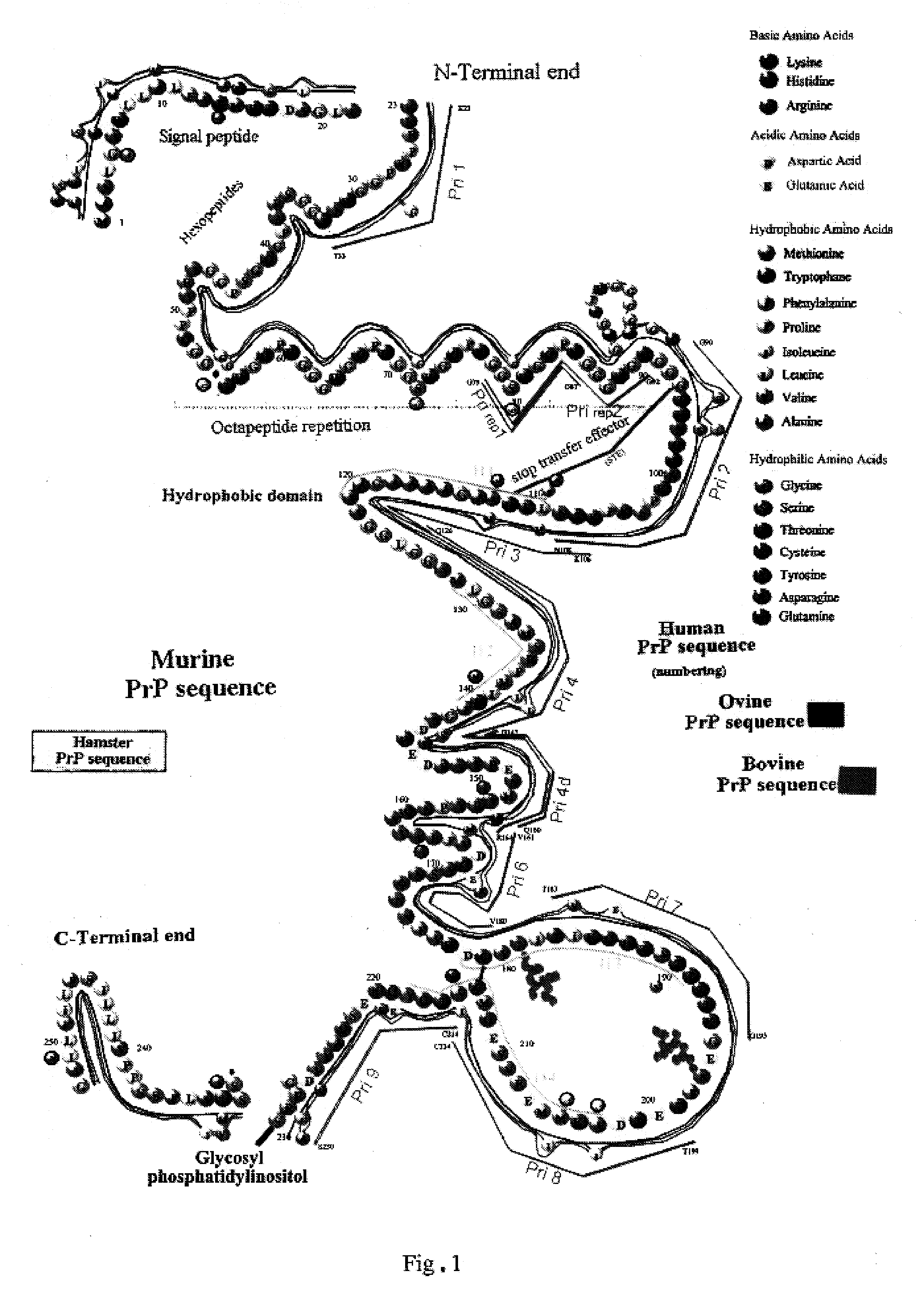
[0087]A peptide representative of the PrP octapeptide motif repeat, for example the motif G-G-W-G-Q-P-H-G-G-G-W-G-Q-G-(NH2), corresponding to sequence 79–92 of the human PrP, was synthesized using an automatic synthesizer (Milligen 9050, Waters, Milford, Mich.). The peptide was covalently coupled to acetylcholinesterase (AchE) via a heterobifunctional reagent, succinimidyl 4-(N-male-imidomethyl)cyclohexane-1-carboxlate (SMCC, Calbiochem, France), as described previously for other peptides or proteins (McLaughlin et al., 1987, Grassi et al., 1989). This method involves reacting a thiol group introduced into the peptide with the maleimide function which was attached to the AchE by reaction with the SMCC. The thiol group was introduced into the peptide by reaction with N-succinimidyl S-acetylthioacetate (SATA) as described previously (McLaughlin et al.,...

example 2

Detection of PrP-res by Western Blotting

I: Treatment of the Sample

(i) Preparation of a Tissue Homogenate from the Various Biological Samples

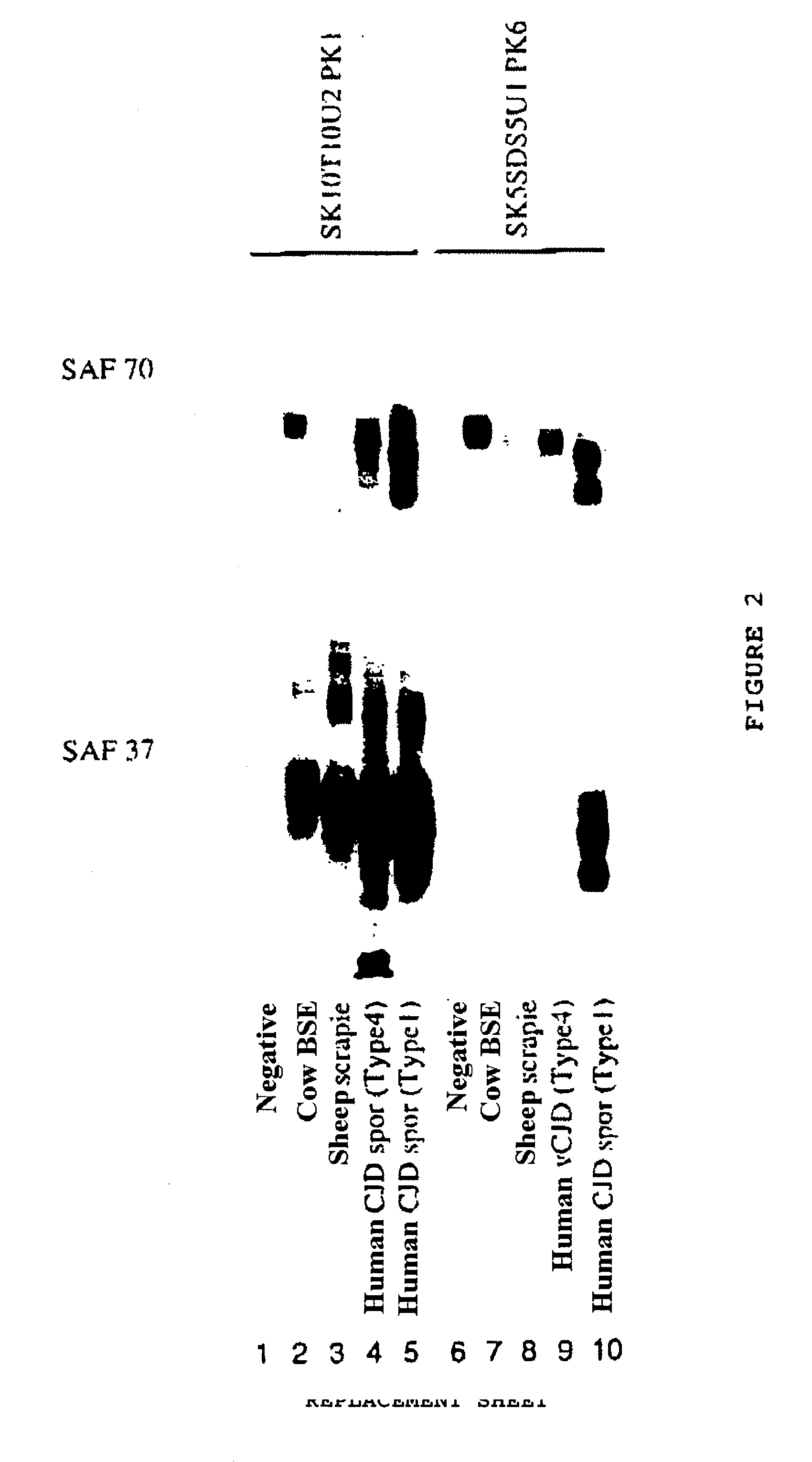
[0091]cow BSE, sheep scrapie, human vCJD (type 4), human sporadic CJD (type 1) and corresponding negative controls

[0092]350 mg of bovine brain are taken: it is ground and homogenized at 20% (w / v) in a 5% glucose solution.

[0093]To perform the homogenization, the brain sample (350 mg) and 1.4 ml of glucose solution are introduced into tubes comprising ceramic beads, and vigorously agitated (Hybaid Ribolyser).

[0094]The positive samples were diluted in a homogenate originating from healthy brains of the corresponding species, as follows:

[0095]for sheep: to 1 / 100th

[0096]for cows: to 1 / 50th

[0097]for humans: type 1 or 4: to 1 / 40th; type 3: to 1 / 80th; type 2: to 1 / 20th

(ii) Conditions for Step (I)

[0098]A first fraction (500 μl) of homogenate at 20% obtained in (i) is incubated with 500 μl of a buffer comprising 10% sarkosyl (SK10), 10% Triton X100 (T10),...

example 3

Detection of the PrP-res with a Two-Site Immunometric Assay Using, as the Capture Antibody, a Monoclonal Antibody which Recognizes the Octapeptide Motif Repeats

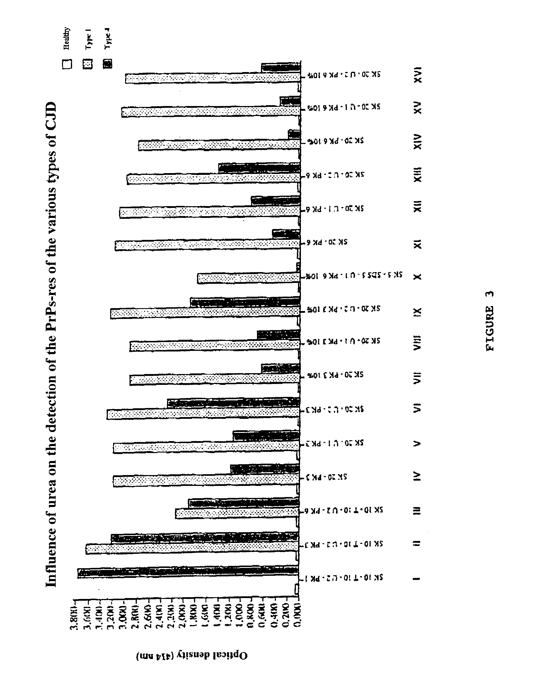
[0119]To carry out a two-site immunometric assay, the pellet obtained in (iv) in example 2 is, for example, dissolved in a buffer comprising sarkosyl (0.25–1%) and urea (0.25–8 M) or SDS (0.25–1%) and urea (0.25–1 M); the sample obtained will preferably be diluted (to ¼ or to ½), after heating, with a buffer containing albumin, producing a final albumin concentration of between 0.1 and 1% (w / v), or with a buffer containing 1% deoxycholate.

[0120]The two-site immunometric assay is performed in microtitration plates containing an antibody which has been immobilized under the conditions already described for other proteins (Grassi et al., 1989). The principle thereof is as follows: the PrP analyzed is recognized by an antibody attached to the solid support (capture antibody) and by a second antibody, which recognizes another part...


PUM

PropertyMeasurementUnit
Temperatureaaaaaaaaaa
Fractionaaaaaaaaaa

Abstract

The invention relates to a method for diagnosing a transmissible spongiform subacute encephalopathy (TSSE) caused by an unconventional transmissible agent (UTA) or prion. The method involves treating a sample suspected of containing a prion with proteinase K for a time and under conditions that completely degrade normal prion protein (Prp-sen), but which only partially digest abnormal prion protein (PrP-res) so that all or some of the octapeptide motif repeats comprising P(H / Q)GGG(- / T)WGQ (SEQ ID NO: 1) in the abnormal prion protein (Prp-res) are retained.

Description

BACKGROUND OF THE INVENTION[0001]1. Field of the Invention[0002]The present invention relates to a method for diagnosing a TSSE caused by a UTA strain, in a biological sample, by detecting PrP-res, and also to the use thereof in the context of differential diagnosis of the various UTA strains in a biological sample.[0003]2. Description of Background Art[0004]Transmissible spongiform subacute encephalopathies (TSSEs) are caused by unconventional transmissible agents (UTAs), also called prions, the precise nature of which to date remains disputed. TSSEs essentially comprise Creutzfeldt-Jakob disease in humans (CJD), scrapie in sheep and goats, and bovine spongiform encephalopathy (BSE) in bovines. Other encephalopathies have been demonstrated in the Felidae, in mink or certain wild animals, such as deer or elk.[0005]These diseases are always fatal and, at the current time, there is no effective treatment.[0006]In TSSEs, there is an accumulation of a host's protein, PrP (or prion prote...

Claims


Application Information

Patent Timeline
IPC IPC(8): C12Q1/37A01N37/18A61K38/00G01N33/566G01N33/53G01N33/531G01N33/68
CPCG01N33/6896G01N2800/2828
Inventor DESLYS, JEAN-PHILIPPECOMOY, EMMANUELGRASSI, JACQUES
Owner COMMISSARIAT A LENERGIE ATOMIQUE ET AUX ENERGIES ALTERNATIVES
Learn More