
Microfluidic apparatus for performing gel protein extractions and methods for using the apparatus

a gel protein and microfluidic technology, applied in the direction of fluid speed measurement, diaphragm, electrolysis, etc., can solve the problems of less distinct protein spots, poor reproducibility, and time-consuming procedures, and achieves rapid and effective protein transfer, large potential drop, and high electric field strength

Inactive Publication Date: 2006-07-04
UNIV OF MARYLAND BALTIMORE +1
View PDF23 Cites 25 Cited by
  • Summary
  • Abstract
  • Description
  • Claims
  • Application Information

AI Technical Summary

Benefits of technology

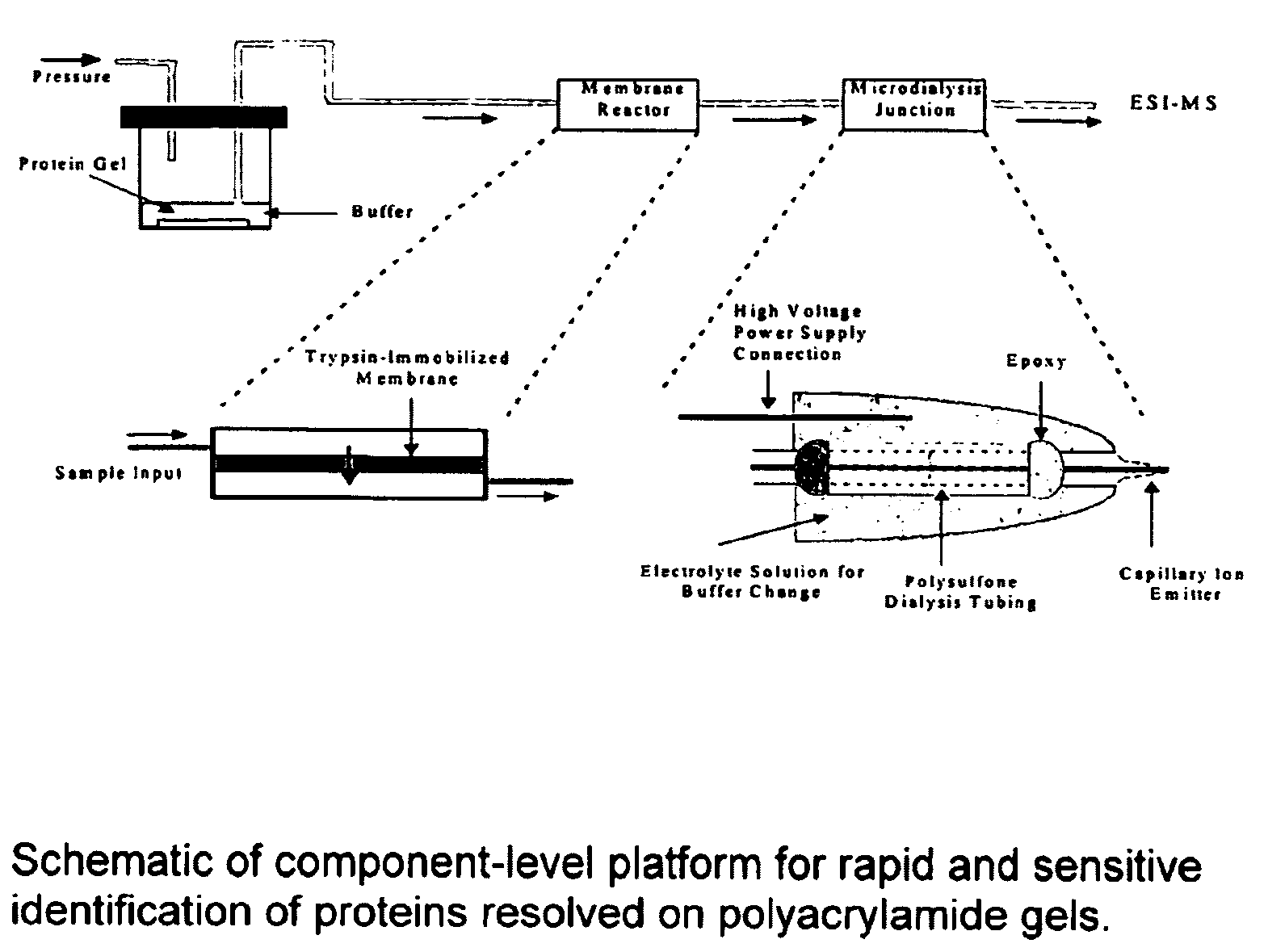
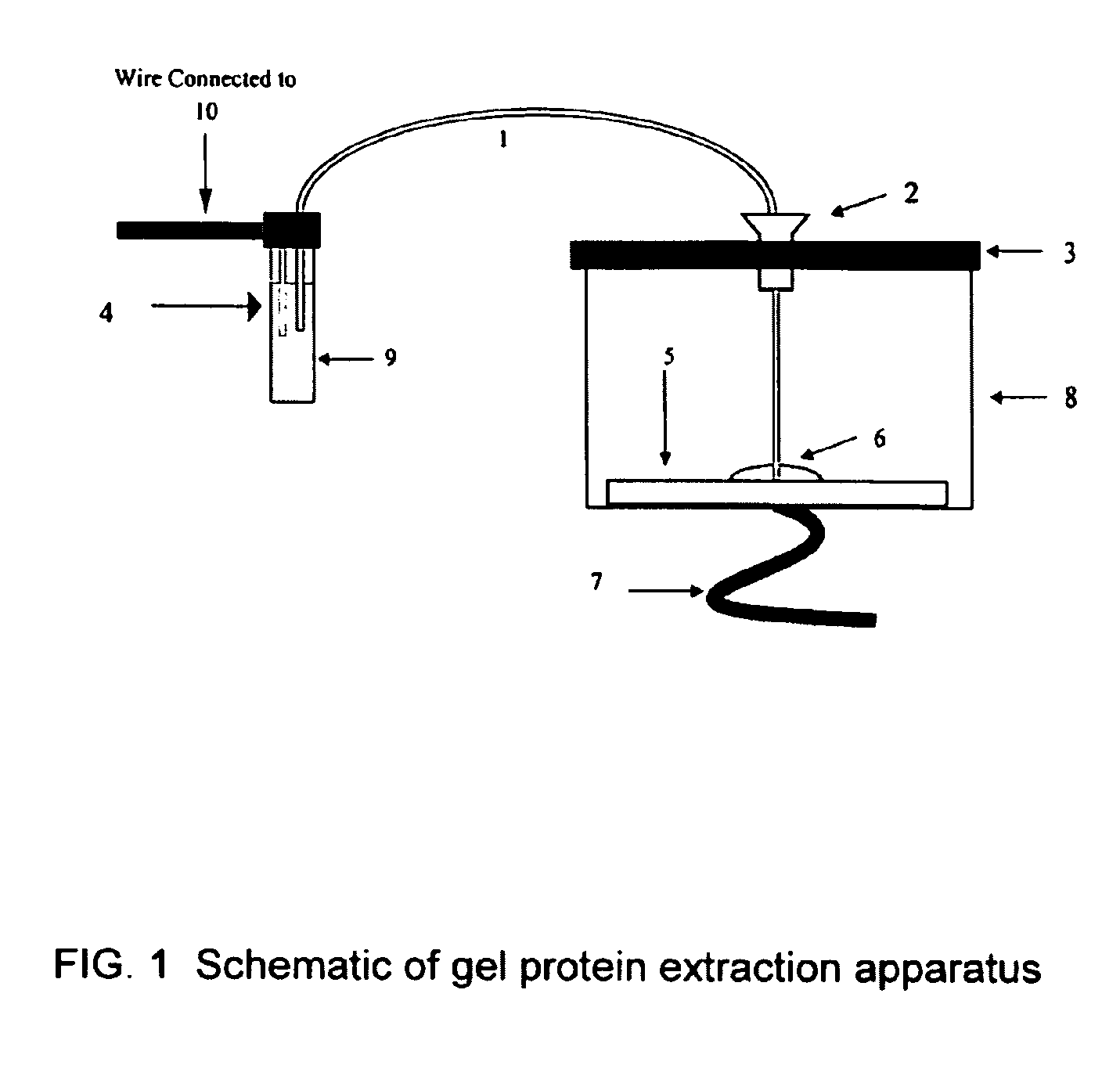
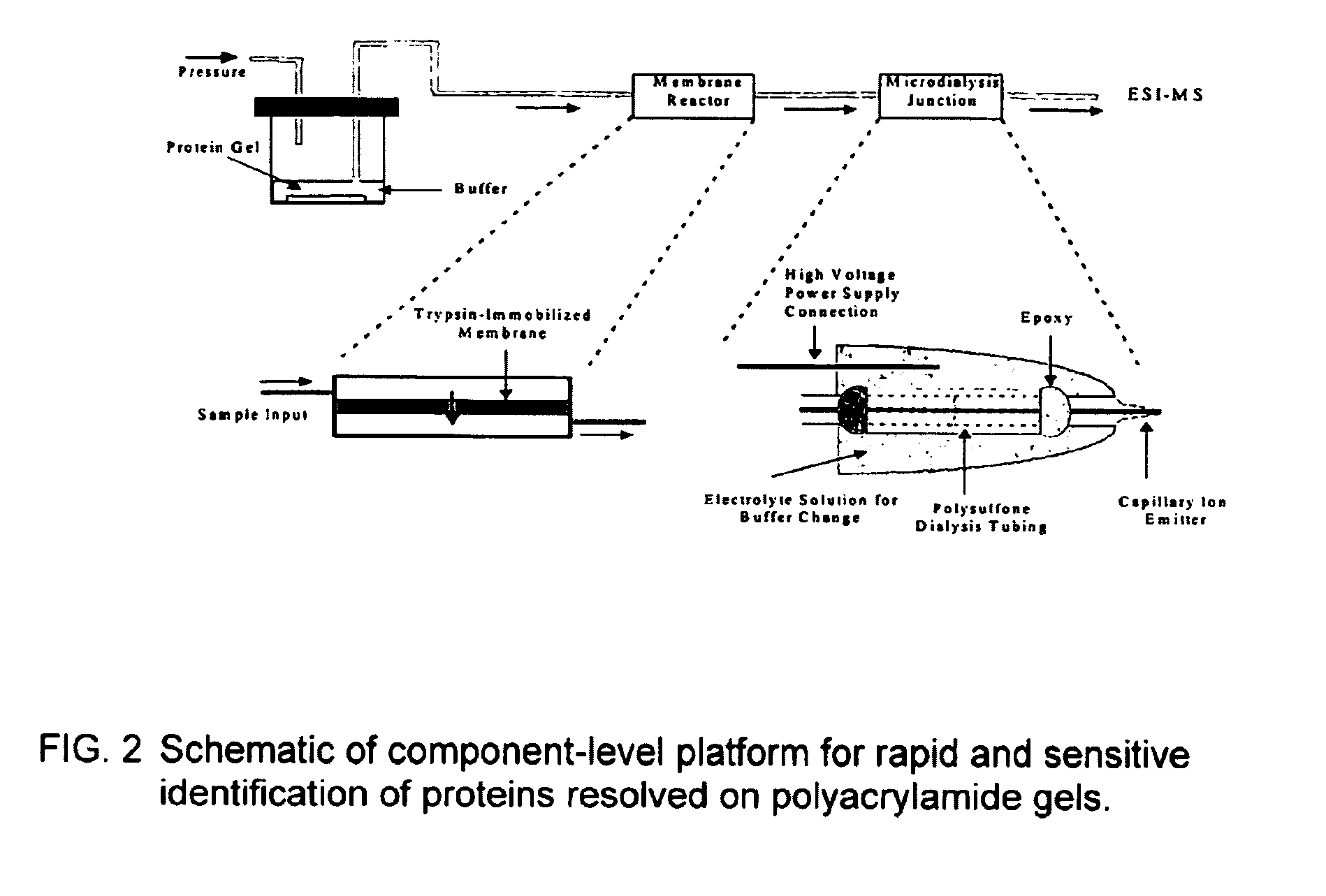
[0012]The invention is directed to a microfluidic apparatus and methods for performing gel protein extractions using the apparatus. The invention involves rapid and efficient extraction of protein analytes directly from 2-D gels using their electrophoretic mobilities, or willingness of a molecule to migrate in accordance with its charge under the application of an electric field in a conductive solution based medium. It is possible to sequentially or simultaneously mobilize several proteins out of gel electrophoresis media and into an apparatus with fluidic channels filled with an electrolyte solution under the application of a high electric field. Once inside the fluidic channels, proteins can be transferred to a miniaturized membrane digestion reactor by pressure or electrokinetic means. The peptide fragments from each protein analyte can be further introduce into a mass spectrometer through fluidic components for protein identification and quantification.

Problems solved by technology

All of these procedures are time-consuming tasks prone to sample loss and analyte dilution.
Attempts to perform native 2-D electrophoresis result in 2-D protein patterns with poor reproducibility, smears, and less distinct protein spots (7).
However, 2-D PAGE is a relatively slow, labor intensive, and cumbersome technology.
Concomitant sensitivity limitations are introduced by the necessary sample handling.
Moreover, 2-D PAGE results from different laboratories can be difficult to compare, and sensitivity is limited by the amount of a protein needed to visualize a spot, typically in the low-nanogram range for silver staining.
A recent study (11), has demonstrated a disadvantage of 2-D PAGE in that only the higher abundance proteins were identified by the 2-D PAGE-MS strategies.
The disadvantage of these strategies, however, is that they all require much larger amounts of protein along with many additional 2-D PAGE and chromatography separations, and therefore may be impractical for studies of small cell populations or tissue samples.
However, none of these disclose methods of increasing the speed of transfer, or supply methods of concentrating the transferred proteins.
Still, the limiting factor for linking 2-D PAGE with mass spectrometric analysis lies in the effective and rapid recovery of peptides, in particular those from low-abundance proteins, from in-gel digestion as well as the extraction and the transfer of gel proteins for sequential or parallel proteolytic digestion (9,58,59).

Method used


Image

  • Microfluidic apparatus for performing gel protein extractions and methods for using the apparatus
  • Microfluidic apparatus for performing gel protein extractions and methods for using the apparatus
  • Microfluidic apparatus for performing gel protein extractions and methods for using the apparatus

Examples


example 1

[0100]As shown in FIG. 3, a protein loading of 100 ng of SDS-cytochrome C complex is extracted from polyacrylamide gel and monitored by UV absorbance at 280 nm. The results are compared with capillary zone electrophoresis (CZE) of protein sample containing SDS-cytochrome C complex with a concentration of 0.5 mg / ml. Thus, the concentration of extracted SDS-cytochrome C complex is estimated to be around 0.25 mg / ml inside a solution plug of 50-100 nL. The migration time of extracted cytochrome C complex to reach the UV detector is approximately 7.4 minutes and slightly longer than that obtained from CZE. UV absorbance detection scarcely reveals the presence of coomassie blue at 280 nm, while at 214 nm a broad peak is noticed prior to protein complexes.

[0101]Based on our estimations regarding the concentration (0.25 mg / ml) and the volume (50-100 nL) of extracted cytochrome C complex, approximately 12.5-25% of cytochrome C from a protein loading of 100 ng is recovered within 2 minutes of...

example 2

[0102]The results summarized in FIG. 5 further demonstrate the ability to rapidly transfer the SDS-cytochrome C complex over a wide range of protein loading, from 5 μg to 50 ng. Peak height of extracted protein complexes decreases with decreasing protein loading on polyacrylamide gel. However, the extent of decrease in peak height is reduced at low protein loadings as the size of a protein spot shrinks with decreased protein loading. The results also demonstrate the ability to extract higher molecular weight proteins such as ovalbumin and β-galactosidase with molecular mass around 45 and 116 kDa, respectively. The peak heights of extracted ovalbumin and β-galactosidase from a gradient gel are about half of those measured from cytochrome C at various protein loadings. However, the UV absorbance of denatured cytochrome C measured at 280 nm is two and four times of those obtained from β-galactosidase and ovalbumin at the same weight concentration, respectively.

example 3

[0103]Denatured and reconstituted cytochrome C at a concentration of 1 mg / ml is introduced into a μ-trypsin membrane reactor at sample flow rates of 0.3, 0.2, and 0.1 μl / min. The corresponding digestion times inside the membrane reactor are 3, 5, 10 minutes, respectively. At room temperature, various degrees of the cytochrome C digestion are observed from partial digestion at 0.3 μl / min to near complete digestion at 0.1 μ / min (see FIGS. 6A, 6B and 6C). Presence of the cytochrome C envelope is quite obvious at a flow rate of 0.3 μl / min. In comparison with solution-based trypsin digestion, the membrane digestion is at least 500-1000 times faster than solution digestion and exhibits the advantage of avoiding autolytic interference from the proteolytic enzyme in the mass spectra.

[0104]To investigate the effect of reaction temperature on protei digestion, the membrane reactor is placed on top of a hot plate. The ambient temperature surrounding the membrane reactor is increased to 40 and ...


PUM

PropertyMeasurementUnit
Fractionaaaaaaaaaa
Fractionaaaaaaaaaa
Thicknessaaaaaaaaaa

Abstract

The invention relates to an apparatus for performing gel protein extractions and methods of using the apparatus.

Description

RELATED APPLICATIONS[0001]This application claims priority from U.S. Ser. No. 60 / 261,215, filed Jan. 16, 2001, and U.S. Ser. No. 60 / 287,754, filed May 1, 2001, which are incorporated by reference in their entirety.FIELD OF THE INVENTION[0002]The invention relates to an apparatus for performing gel protein extractions and methods of using the apparatus.BACKGROUND OF THE INVENTION[0003]Complex biological processes, including development, differentiation, and signal transduction, involve the coordinated expression of multiple genes and proteins, as well as control of their function. The identification and quantification of multiple proteins that constitute and control a particular process is important for understanding the regulation of biological systems. Additionally, the ability to monitor the presence or absence of particular proteins, an increase or decrease in protein expression, a change in protein microheterogeneity, or a combination of these modifications may be used for early...

Claims


Application Information

Patent Timeline
IPC IPC(8): G01N27/447G01N27/453B81B1/00
CPCG01N27/4473G01N27/44791G01N27/44773
Inventor LEE, CHENGDEVOE, DON
Owner UNIV OF MARYLAND BALTIMORE
Learn More