Mammography film and imaging assembly for use with rhodium or tungsten anodes

Inactive Publication Date: 2005-03-08
CARESTREAM HEALTH INC
View PDF25 Cites 0 Cited by
  • Summary
  • Abstract
  • Description
  • Claims
  • Application Information

AI Technical Summary

Benefits of technology

The present invention provides a means for providing radiographic images for mammography unexpectedly exhibiting improved image quality while minimizing radiation dosage to which patients are exposed. In particular, image quality can be improved with the present invention by increasing image contrast, decreasing “noise” (for example, film granularity), or both. These advantages are possible with the unique radiographic film and imaging assembly of this invention and thereby allow patient imaging to be carried out using imaging equipment containing rhodium or tungsten anodes without sacrificing image quality such as image contrast.
In addition, all other desirable sensitometric properties are maintained and the radiographic film can be rapidly processed in the same conventional processing equipment and compositions.

Problems solved by technology

This can contribute to loss of image sharpness and contrast.
Thus mammography is a very difficult task in medical radiography.
Rhodium anodes are also known in the art for lowering patient exposure to radiation, but in the case of mammography, poorer image quality is usually the result when they are used.

Method used


Image

  • Mammography film and imaging assembly for use with rhodium or tungsten anodes
  • Mammography film and imaging assembly for use with rhodium or tungsten anodes
  • Mammography film and imaging assembly for use with rhodium or tungsten anodes

Examples


example

Radiographic Film A (Control)

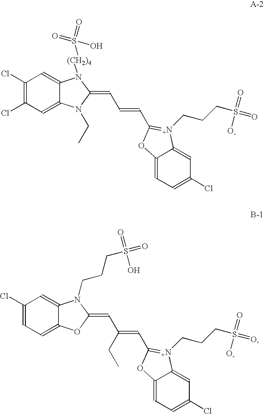
Radiographic Film A was a single-coated film having the a silver halide emulsion on one side of a blue-tinted 170 μm transparent poly(ethylene terephthalate) film support and a pelloid layer on the opposite side. The emulsion was chemically sensitized with sulfur and gold and spectrally sensitized with the following dye A-1:

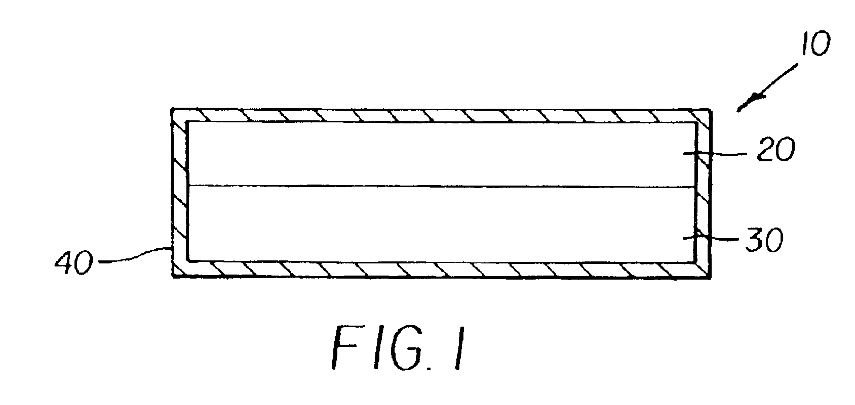
Radiographic Film A had the following layer arrangement:OvercoatInterlayerEmulsion LayerSupportPelloid LayerOvercoat

The noted layers were prepared from the following formulations.

Coverage (mg / dm2)Overcoat FormulationGelatin vehicle4.4Methyl methacrylate matte beads0.35Carboxymethyl casein0.73Colloidal silica (LUDOX AM)1.1Polyacrylamide0.85Chrome alum0.032Resorcinol0.073Dow Corning Silicone0.153TRITON X-200 surfactant (Union Carbide)0.26LODYNE S-100 surfactant (Ciba Specialty Chem.)0.0097Interlayer FormulationGelatin vehicle4.4Emulsion Layer FormulationCubic grain emulsion51.1[AgBr 0.85 μm average size]Gelatin vehicle34.9Spectral sens...


PUM


Abstract

A radiographic silver halide film comprises a cubic grain silver halide emulsion layer on one side of the support and a tabular grain silver halide emulsion layer on the other side. The cubic grain silver halide emulsion layer comprises a combination of first and second spectral sensitizing dyes that provides a combined maximum J-aggregate absorption on the cubic silver halide grains of from about 540 to about 560 nm. The first spectral sensitizing dye is an anionic benzimidazole-benzoxazole carbocyanine, the second spectral sensitizing dye is an anionic oxycarbocyanine. The cubic grain silver halide emulsion layer also includes a mixture of gelatin or a gelatin derivative and a second hydrophilic binder other than gelatin or a gelatin derivative at a weight ratio of first to second hydrophilic binder of from about 2:1 to about 5:1. The cubic silver halide grains comprise from about 1 to about 20 mol % chloride and from about 0.25 to about 1.5 mol % iodide, which cubic silver halide grains have an average ECD of from about 0.65 to about 0.8 μm. Moreover, the cubic silver halide grains are doped with a hexacoordination complex compound within part or all of the innermost 95% of the grains. These films can be exposed to provide a black-and-white image having a d(γ) / d(log E) value greater than 5 and are particularly useful when imaged using X-radiation generating devices comprising rhodium or tungsten anodes.

Description

FIELD OF THE INVENTIONThis invention is directed to radiography. In particular, it is directed to a radiographic silver halide film and imaging assembly that are useful for providing medical diagnostic images of soft tissues such as in mammography. These films provide advantages when exposed to X-radiation generated using rhodium or tungsten anodes.BACKGROUND OF THE INVENTIONThe use of radiation-sensitive silver halide emulsions for medical diagnostic imaging can be traced to Roentgen's discovery of X-radiation by the inadvertent exposure of a silver halide film. Eastman Kodak Company then introduced its first product specifically that was intended to be exposed by X-radiation in 1913.In conventional medical diagnostic imaging the object is to obtain an image of a patient's internal anatomy with as little X-radiation exposure as possible. The fastest imaging speeds are realized by mounting a dual-coated radiographic element between a pair of fluorescent intensifying screens for imag...

Claims


Application Information

Patent Timeline
IPC IPC(8): G03C5/16G03C5/17G03C1/08G03C1/04G03C1/14G03C1/29G03C1/46G03C1/18G03C5/26
CPCG03C5/16G03C5/17G03C1/04G03C1/08G03C1/18G03C1/29Y10S430/167G03C5/26G03C1/46G03C2001/03541G03C2200/58G03C2001/03594G03C1/0051G03C2200/52
Inventor DICKERSON, ROBERT E.MOORE, WILLIAM E.STEKLENSKI, DAVID J.
Owner CARESTREAM HEALTH INC
Why Eureka
Try Eureka