Systems and methods for assessing specimen fixation duration and quality using vibrational spectroscopy

a vibration spectroscopy and specimen technology, applied in the field of systems and methods for assessing specimen fixation duration and quality using vibration spectroscopy, can solve problems such as inconclusive results and/or misdiagnosis

Pending Publication Date: 2022-05-05
VENTANA MEDICAL SYST INC
View PDF0 Cites 2 Cited by
  • Summary
  • Abstract
  • Description
  • Claims
  • Application Information

AI Technical Summary

Benefits of technology

The patent text describes a system and method that can accurately predict the fixation status of a biological specimen. This means that the system can determine if the specimen has been fixed for a long time or if it is still unfixed. This is important because the fixation process can affect the quality of the experiment and the results need to be reliable. The system can also estimate the duration of the fixation process, which is important for understanding the effects of the fixation process on the biological specimen. Overall, the patent describes a system that can accurately and in detail predict the fixation status of a biological specimen.

Problems solved by technology

Variations in the fixation of a biological specimen, e.g. a tissue sample, may impact downstream labeling and / or staining processes, which may result in inconclusive results and / or a misdiagnosis.

Method used


Image

  • Systems and methods for assessing specimen fixation duration and quality using vibrational spectroscopy
  • Systems and methods for assessing specimen fixation duration and quality using vibrational spectroscopy
  • Systems and methods for assessing specimen fixation duration and quality using vibrational spectroscopy

Examples


example 1

g Fixation Duration Using a Trained Fixation Estimation Engine

[0153]The Example provided below is illustrative of the methods described herein.

[0154]Introduction

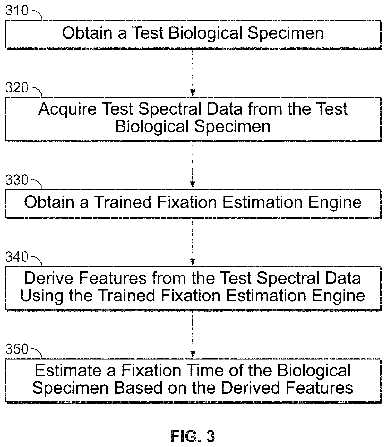
[0155]Modern histology is built on the 100+year old cornerstone technology of formaldehyde fixation which preserves tissue's biostructure by arresting mechanisms of molecular degradation by crosslinking a tissue's biostructure. How thoroughly a sample is fixed can significantly impact detected protein expression and in extreme cases a patient's diagnosis. For example, ASCO / CAP guidelines require samples be fixed for 6-72 hours. Currently tissue fixation in clinical practice is laboratory specific and highly variable. Despite its importance, there are however no analytical methods of measuring fixation quality.

[0156]Mid infrared spectroscopy (mid-IR) is a powerful optical technique that probes the vibrational state of individual molecules in the tissue and is very sensitive to the conformational state of proteins. This extrem...

example 2

a Fixation Estimation Engine

[0186]In one embodiment, a projection-onto-latent-structure (also partial least squares regression) PLSR model is trained using spectra acquired from a mid-IR microscope. The mid-IR spectra is collected at roughly one hundred locations throughout the tissue, so the average spectrum is representative of the average of the tissue. All spectra were atmospherically corrected to remove CO2 contamination, baseline-corrected using concave rubber band correction with 10 iterations and 64 baseline points, then spectra were amplitude normalized. Finally, all the spectra from each tissue were averaged together. Next a PLSR regression model was trained by splitting the data into 5 parts, meaning every 5th spectrum was pulled out of the data set and only used exclusively for validation of the model. About 80% of the tissue's spectra was then used to train a PLSR model using 2-fold cross validation and the raw absorption spectra (as opposed to the 1st, 2nd, etc. deriva...


PUM


Abstract

The present disclosure relates to automated systems (200) and methods for quantitatively determining a fixation duration of a biological specimen using a trained fixation estimation engine (210). In some embodiments, the trained fixation estimation (210) engine includes a neural network. In some embodiments, the trained fixation estimation (210) engine includes a supervised classifier.

Description

CROSS-REFERENCE TO RELATED APPLICATIONS[0001]The present application is a continuation of International Application No. PCT / EP2020 / 073787, which application claims the benefit of the filing date of U.S. Patent Application No. 62 / 892,678 filed on Aug. 28, 2019, the disclosures of which are hereby incorporated by reference herein in their entireties.BACKGROUND OF THE DISCLOSURE[0002]Immunohistochemical (IHC) slide staining can be utilized to identify proteins in cells of a tissue section and hence is widely used in the study of different types of cells, such as cancerous cells and immune cells in biological tissue. Thus, IHC staining may be used in research to understand the distribution and localization of the differentially expressed biomarkers of immune cells (such as T-cells or B-cells) in a cancerous tissue for an immune response study. For example, tumors often contain infiltrates of immune cells, which may prevent the development of tumors or favor the outgrowth of tumors.[0003...

Claims


Application Information

Patent Timeline
IPC IPC(8): G01N21/65G01N21/3563G01N21/39G01N33/483G06N3/08
CPCG01N21/65G01N21/3563G01N2201/06113G01N33/4833G06N3/08G01N21/39G01N21/35G01N2201/1296G01N2201/1293G01N2021/399G01N1/30G01N21/1702
Inventor BAUER, DANIELCHAFIN, DAVID
Owner VENTANA MEDICAL SYST INC
Why Eureka
Try Eureka