
Hyaluronic Acid Binding Domain-Growth Factor Fusion Protein cDNAs and Fusion Proteins for Cartilage Matrix Preservation and Report

a technology of hyaluronic acid and growth factor, which is applied in the direction of fusions for specific cell targeting, hormone peptides, genetic material ingredients, etc., can solve the problems of limited factors, unsolved clinical problems for articular cartilage repair, and damage to articular cartilage, so as to increase the responsiveness rate of chondrocytes, and reduce gf removal rate

Inactive Publication Date: 2020-02-06
INDIANA UNIVERISY RES & TECH CORP +1
View PDF0 Cites 1 Cited by
  • Summary
  • Abstract
  • Description
  • Claims
  • Application Information

AI Technical Summary

Benefits of technology

[0070]Also provided are methods to increase responsiveness rates of arthritic chondrocytes to GF in a subject, comprising expressing a nucleic acid molecule herein in at least one chondrocyte in at least one arthritic joint of a subject and increasing responsiveness rates of arthritic chondrocytes to GF in the subject.
[0076]Also provided are methods to increase sports performance in an athlete, comprising expressing a nucleic acid molecule herein in at least one joint or intervertebral disc of an athlete and increasing sports performance in the athlete.
[0077]Also provided are methods herein, wherein the sports performance is selected from the group consisting of: increased speed; increased endurance; increased weight-lifting ability; increased flexibility; increased strength; increased resistance to impact; increased concentration; and increased career length.
[0085]Also provided are methods to increase responsiveness rates of chondrocytes to GF in a subject, comprising introducing a fusion protein herein in at least one chondrocyte of a subject and increasing responsiveness rates of chondrocytes to GF in the subject.
[0091]Also provided are methods to increase sports performance in an athlete, comprising introducing a fusion protein herein in at least one joint or intervertebral disc of an athlete and increasing sports performance in the athlete.
[0092]Also provided are methods herein, wherein the sports performance is selected from the group consisting of: increased speed; increased endurance; increased weight-lifting ability; increased flexibility; increased strength; increased resistance to impact; increased concentration; and increased career length.

Problems solved by technology

Articular cartilage damage is a major cause of disability in the form of arthritis and joint trauma.
Because articular cartilage lacks the ability to effectively repair itself, articular cartilage repair is an unsolved clinical problem.
When delivered to articular cartilage as potential therapeutic agents, these factors are limited by rapid elimination from the joint, slower uptake by the articular cartilage containing the target cells, and, when present in the cartilage or repair tissue, are subject to diffusion out of the tissue where it is needed.
However, an unpublished clinical trial of IGF-1 delivery to human knee joints was evidently unsuccessful.
A major limitation of current approaches to IGF-1 therapy include: 1) The rapid removal of IGF-1 from the joint through the synovium, 2) the relatively slow diffusion of IGF-1 into the articular cartilage where it can act on the cells, and 3) limited responsiveness of arthritic chondrocytes to the IGF-1.
Other limitations of the related art will become apparent to those of skill in the art upon a reading of the specification and a study of the drawings.

Method used


Image

  • Hyaluronic Acid Binding Domain-Growth Factor Fusion Protein cDNAs and Fusion Proteins for Cartilage Matrix Preservation and Report
  • Hyaluronic Acid Binding Domain-Growth Factor Fusion Protein cDNAs and Fusion Proteins for Cartilage Matrix Preservation and Report
  • Hyaluronic Acid Binding Domain-Growth Factor Fusion Protein cDNAs and Fusion Proteins for Cartilage Matrix Preservation and Report

Examples


example 1

and Methods

[0124]Fusion Proteins

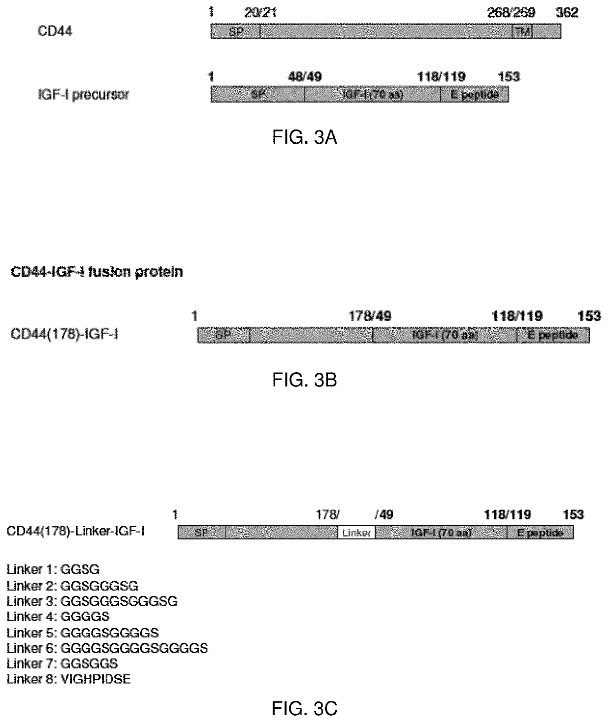
[0125]The nucleic acid sequences encoding the described proteins and chimeric nucleic acid sequences encoding fusion proteins may be constructed. The chimeric nucleic acid molecules may be prepared with sections encoding a linking peptide connecting the protein portions of the encoded fusion protein. Optionally, the peptide linker may be selected to include residues imparting stearic flexibility in order to enhance the function of the fusion protein.

[0126]Fusion Protein Linkers

[0127]The components of the fusion proteins may be operatively linked directly one to the other. The two protein components of the fusion protein may be directly linked via an amino terminus to carboxy terminus peptide bond.

[0128]Alternatively, one or more linker molecules connect the two portions of the fusion protein. The linker molecule allows the two portions of the fusion protein increased stearic freedom. In one embodiment, the linkers are peptide linkers. Peptides for lin...

example 2

of Hyaluronic Acid Binding—Insulin-Like Growth Factor I Fusion Protein cDNA Constructs

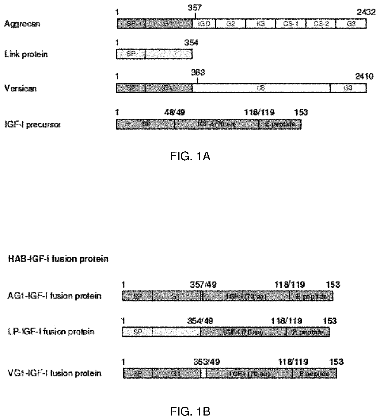
[0158]Three cDNAs were created, encoding three hyaluronic acid-binding domain (HAB)—insulin-like growth factor I (IGF-I) fusion proteins (HAB-IGF-I) by coupling the sequence encoding IGF-I with those encoding a hyaluronic acid binding region derived from three cartilage matrix proteins. These regions include 1) the aggrecan G1 domain (AG1), 2) the versican G1 domain (VG1) and 3) link protein (LP). The resulting fusion proteins are designated AG1-IGF-I, VG1-IGF-I, and LP-IGF-I respectively (FIG. 1B). Further, the encoded fusion proteins were created by transferring the cDNAs into producer cells and the selected therapeutic target cells, articular chondrocytes.

[0159]To facilitate the HAB-IGF-I constructs to be used for human therapeutic purposes, the DNA constructs encoding all the HAB-IGF-I fusion proteins were generated from native human matrix protein gene sequences and the native human IGF-I gene...

example 3

n of Fusion Proteins and Demonstration of Fusion Protein Integrity

[0162]To promote articular cartilage preservation or repair using these fusion proteins as therapeutic agents, it is necessary to have a production source for the proteins. A method of production employs the fusion protein-encoding cDNAs described above and a non-viral (plasmid) vector that the inventors developed previously, Shi et al., Effect of Transfection Strategy on Growth Factor Overexpression by Articular Chondrocytes, J. of Orthopaedic Research, January 2010, hereby incorporated by reference. The cDNAs were inserted into the vector and used to transfect HEK 293 cells (Stratagene). The HEK 293 cells then use these inserted transgenes to synthesize the HAB-IGF-I fusion proteins. On day 3 after transfection conditioned medium was collected for measurement of HAB-IGF-I fusion proteins produced as transgene products by the HEK 293 cells. The production and integrity of these proteins was confirmed by IGF-I ELISA (...


PUM

PropertyMeasurementUnit
volumesaaaaaaaaaa
volumeaaaaaaaaaa
volumeaaaaaaaaaa

Abstract

The present invention provides isolated nucleic acid compounds, amino acids compounds and related materials, along with methods to make and use the compounds. In particular, there are provided gene therapy materials useful for inducing positive physiological responses in tissues, and fusion proteins encoded by the gene therapy materials.

Description

CROSS-REFERENCE TO RELATED APPLICATIONS[0001]This application is a Continuation Application of U.S. application Ser. No. 14 / 294,305 filed Jun. 3, 2014 which claims priority to U.S. Provisional Application No. 61 / 830,925, filed on Jun. 4, 2013, the entire disclosure of which is expressly incorporated herein by reference for all purposes.STATEMENT REGARDING FEDERALLY SPONSORED RESEARCH[0002]This invention was made with government support under AR047702 awarded by the National Institutes of Health. The government has certain rights in this invention.SEQUENCE LISTING[0003]The Sequence Listing, filed electronically in ASCII text format and identified as 3619_54880_SEQ_LIST_IURTC-11114.txt, was created on May 29, 2014, is 140,103 bytes in size and is hereby incorporated by reference.BACKGROUND OF THE INVENTION[0004]Articular cartilage damage is a major cause of disability in the form of arthritis and joint trauma. Because articular cartilage lacks the ability to effectively repair itself,...

Claims


Application Information

Patent Timeline
Patent Type & Authority Applications(United States)
IPC IPC(8): A61K48/00C07K14/65
CPCC07K14/65A61K48/0058C12N2750/14143C07K2319/20C07K2319/33C07K14/78
Inventor TRIPPEL, STEPHEN B.SHI, SHUILIANG
Owner INDIANA UNIVERISY RES & TECH CORP
Learn More