
Differential diagnosis

a diagnostic and differential diagnosis technology, applied in the field of in vivo imaging, can solve problems such as difficulty in committing to an accurate clinical diagnosis

Active Publication Date: 2019-10-31
GE HEALTHCARE LTD +2
View PDF0 Cites 1 Cited by
  • Summary
  • Abstract
  • Description
  • Claims
  • Application Information

AI Technical Summary

Benefits of technology

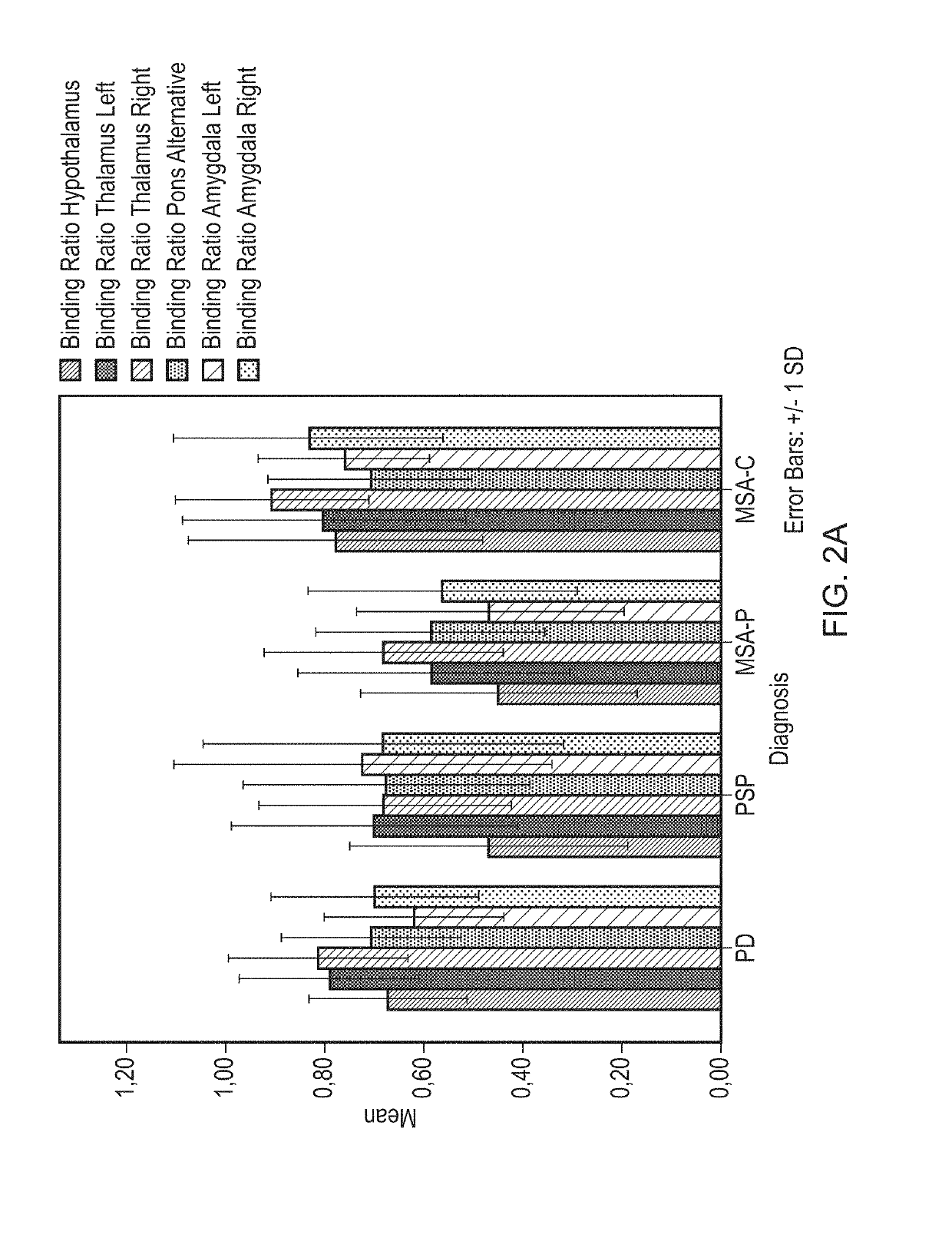
The present invention discusses a method for accurately diagnosing Parkinson's disease (PD) from multiple system atrophy Parkinsonian type (MSA-P) and progressive supranuclear palsy (PSP) using a radiolabelled phenyltropane in vivo imaging agent. The method involves administering the imaging agent to a subject, obtaining an in vivo image of the brain's uptake of the imaging agent, and analyzing the image to determine a binding ratio for selected brain regions. This ratio is then compared with a receiver operating characteristic (ROC) curve to differentiate PD from MSA-P or PSP. The invention provides a reliable tool for accurate diagnosis of these neurological disorders.

Problems solved by technology

The similarity of symptom profile, especially in the early clinical stages of these different Parkinsonian diseases, often makes it difficult to commit to an accurate clinical diagnosis.
However, to make a distinction between PD, MSA-P and PSP, based on DAT imaging as assessed by [123I]FP-CIT SPECT imaging, is more of a challenge.

Method used


Image

  • Differential diagnosis
  • Differential diagnosis
  • Differential diagnosis

Examples


example 1

Imaging

Example 1(i): Acquisition and Reconstruction Procedure

[0072]All patients received oral potassium perchlorate to block thyroid uptake of free radioactive iodide. [123I]FP-CIT was injected intravenously 3 hours before image acquisition at an approximate dose of 185 MBq (specific activity >185 MBq / nmol; radiochemical purity >99%). Subjects were imaged using a dual-head gamma camera (E.Cam; Siemens. Munich. Germany) with a fan-beam collimator. 60×30 second views per head over a 180° orbit on a 128×128 pixel matrix were acquired resulting in a total imaging time of 30 minutes. Image reconstruction was performed using a filtered back projection with a Butterworth filter (order 8. cut-off 0.6 cycles / cm; voxel size: 3.9 mm3 after reconstruction). Scans were reoriented manually to ensure that left and right striatum were aligned.

Example 1(ii): Region-of-Interest (ROI) Analysis

[0073]ROIs were defined for the DAT-rich caudate nucleus and the SERT-rich thalamus from the automated anatomi...

example 1 (

Example 1(vi): ROC Analysis

[0102]ROC-Curve Mean BRs of Every ROI Combined

[0103]To determine a possible combination of striatal with extrastriatal ROIs to increase differential diagnostic possibilities between PD, MSA-P and PSP, receiver operator characteristics curves (ROC-curves) were made for several combinations of ROIs, using the following method:

[0104]For this ROC analysis patients not on SSRIs were used. To test for the possibility to distinguish between PD and non-PD in this group, comparison between groups as PD vs non-PD was determined, the latter entailing MSA-P and PSP.

[0105]Followed Steps:[0106]1. Logistic regression on PD, MSA-P, PSP patients divided in 2 groups→PD (yes / no) with all (10) striatal and extrastriatal BRs.[0107]2. Estimated expected values are saved.[0108]3. Estimated values are combined with PD (yes / no) in ROC curve.

[0109]From the coordinates of the curve Table, the following values were subtracted:

[0110]Sensitivity / specificity (caudate and posterior putam...


PUM

PropertyMeasurementUnit
pHaaaaaaaaaa
timeaaaaaaaaaa
thresholdaaaaaaaaaa

Abstract

The present application provides a means for differential diagnosis of Parkinson's disease and the clinically similar Parkinsonian disorders multiple system atrophy with predominantly Parkinsonian features (MSA-P) and progressive supranuclear palsy (PSP).

Description

TECHNICAL FIELD OF THE INVENTION[0001]The present invention relates to in vivo imaging and in particular in vivo imaging of the brain to distinguish Parkinson's disease (PD) from other Parkinsonian disorders typically having similar clinical presentation.DESCRIPTION OF RELATED ART[0002]PD is a neurodegenerative movement disorder with motor symptoms including bradykinesia, rigidity, postural instability and resting tremor (Gibb & Lees 1988 J Neurol Neurosurg Psych; 51: 745-752). Loss of dopaminergic neurons in the substantia nigra plays a major role in the aetiology of these motor signs. Less prevalent movement disorders are progressive supranuclear palsy (PSP) and multisystem atrophy (MSA). Clinically and neuropathologically, MSA can be divided into MSA with predominantly Parkinsonian (MSA-P) or cerebellar (MSA-C) features. Particularly, MSA-P and PSP patients frequently display overlap of motor symptoms with PD. These disorders are therefore called degenerative Parkinsonisms. The s...

Claims


Application Information

Patent Timeline
Patent Type & Authority Applications(United States)
IPC IPC(8): A61K51/04A61P25/16G16H50/30
CPCA61K51/0448G16H50/30A61P25/16C07B59/002G01N33/6896G01N33/0093
Inventor JONES, PAUL ALEXANDERBOOIJ, JANVRIEND, CHRIS
Owner GE HEALTHCARE LTD
Learn More