Method of inducing cardiomyocytes proliferation and treating heart diseases
a cardiomyocyte and induction technology, applied in the field of inducing cardiomyocyte proliferation and treating heart diseases, can solve the problems that the role of agrin, dystroglycan and their downstream elements has never been studied in the contex
- Summary
- Abstract
- Description
- Claims
- Application Information
AI Technical Summary
Benefits of technology
Problems solved by technology
Method used
Image
Examples
example 1
Materials and Methods
[0180]Isolation of Cardiac Cells
[0181]Primary cardiac cells were isolated from ICR 1-day-old (P1) and 7-day-old (P7) mice using a neonatal dissociation kit (gentleMACS), according to the manufacturer's instructions, and cultured in gelatin-coated (0.02%, G1393, Sigma) wells with DMEM / F12 medium supplemented with L-glutamine, Na-pyruvate, non-essential amino acids, penicillin, streptomycin, 5% horse serum and 10% FBS at 37° C. and 5% CO2. In experiments involving administration of either c-terminal recombinant Agrin (550-AG) (R&D Systems), ECM fragments or broad MMP inhibitors (GM6001, Marimastat), the cells were allowed to adhere for 48 h prior to treatment. Subsequently, the medium was replaced with FBS-free medium containing 5% horse serum and the indicated treatment doses for 72 h. Cells were fixed in 4% paraformaldehyde (PFA) and stained for markers of interest.
[0182]Preparation of Heart Derived ECM
[0183]Hearts were taken from ICR mice (1 and 7 day old), and...
example 2
P1 Cardiac ECM Increases CM Proliferation in a MMP Dependent Manner
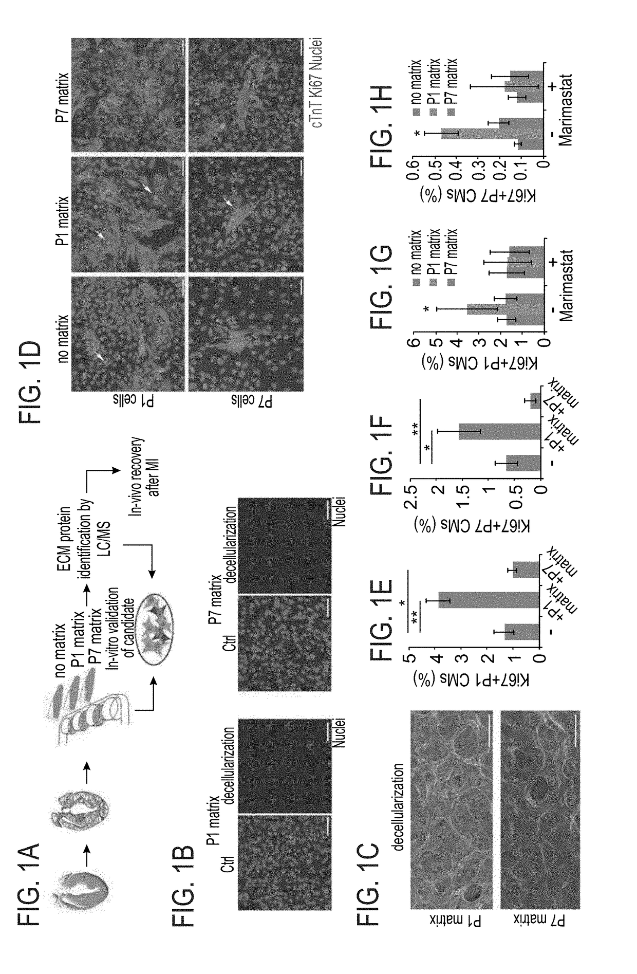
[0200]The effect of the cardiac ECM on CM turnover during the regenerative timeframe in mice was determined {Porrello, 2011 #11}. For that purpose P1 and P7 hearts underwent decellularization (FIG. 1A) to produce cell free ECM fragments as confirmed by DAPI staining and scanning electron microscopy (FIGS. 1B-C). In vitro administration of P1 ECM fragments promoted an increase in both P1 and P7 CM cell-cycle activity, whereas P7 ECM fragments reduced cell cycle re-entry (FIGS. 1D-F).
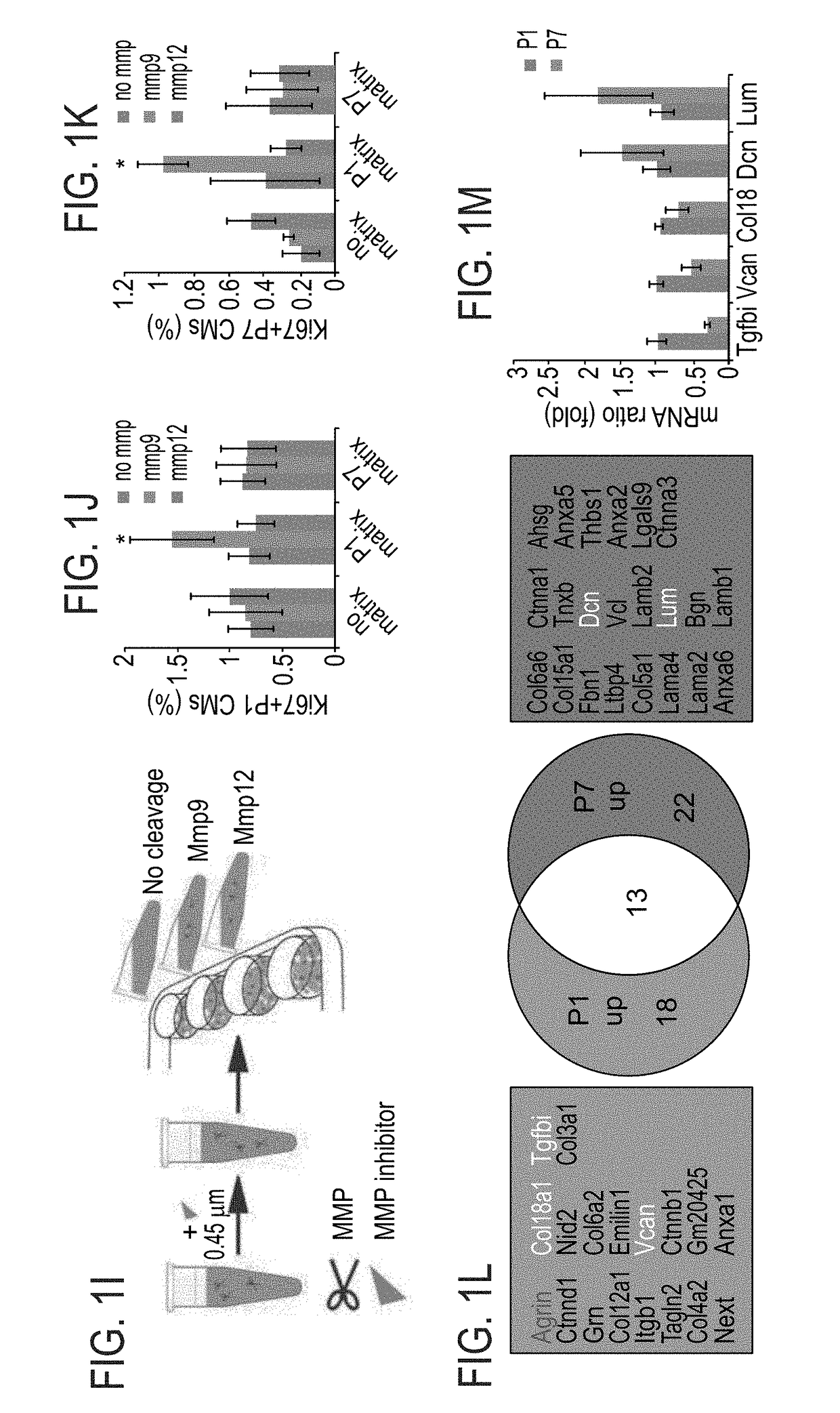
[0201]To gain further insights into the mechanism by which P1 ECM induces CM proliferation, a broad MMP inhibitor (Marimastat) was administered to the culture. Addition of the inhibitor to CM cultures containing ECM fragments derived from P1 hearts abolished the activation of CM proliferation by the P1 ECM explants (FIGS. 1G-H). Addition of the inhibitor to either control cultures or to cultures with ECM fragments derived from P7 hearts, d...
example 3
Endocardial / Endothelial Derived Agrin Promotes CM Proliferation
[0205]The expression levels of Agrin in the heart were then tested {Moll, 2001 #111; McKee, 2009 #112}. Immunofluorescence analysis validated previous finding showing the downregulation of Agrin expression (RNA and protein) at P7 hearts, compared to P1 (FIGS. 2A-C). Next, the cell population which produces Agrin was identified. To do so, P1 cardiac cells were separated to 3 different populations: CMs, fibroblasts (FBs) and endothelial cells (ECs). Enrichment of CMs, FBs and ECs cell populations was confirmed using qPCR for known markers of each cell population (αMHC, CD90 and CD31, respectively, FIG. 2I). Agrin mRNA expression was significantly enriched in the EC population relative to all other cell types (FIG. 2D). The reduction of Agrin expression during the first week of life correlates with the loss of cardiac regenerative response in mice, therefore, it may suggest a role for Agrin during the regeneration process (...
PUM
| Property | Measurement | Unit |
|---|---|---|
| Solubility (mass) | aaaaa | aaaaa |
Abstract
Description
Claims
Application Information
 - R&D
- Intellectual Property
- Life Sciences
- Materials
- Tech Scout
- Unparalleled Data Quality
- Higher Quality Content
- 60% Fewer Hallucinations
Browse by: Latest US Patents, China's latest patents, Technical Efficacy Thesaurus, Application Domain, Technology Topic, Popular Technical Reports.
© 2025 PatSnap. All rights reserved.Legal|Privacy policy|Modern Slavery Act Transparency Statement|Sitemap|About US| Contact US: help@patsnap.com


