
Method for positioning, in cytoplasm, antibody having complete immunoglobulin form by penetrating antibody through cell membrane, and use for same

a technology of cytoplasm and antibody, which is applied in the direction of antibody medical ingredients, instruments, drug compositions, etc., can solve the problems of conventional antibodies that cannot directly penetrate living cells, antibodies and macromolecular bio-drugs, and cannot pass the hydrophobic cell membrane, etc., to inhibit the activity of ras, no cytotoxicity, and stable ability to penetrate the cytosol

Active Publication Date: 2017-08-03
ORUM THERAPEUTICS INC
View PDF0 Cites 8 Cited by
  • Summary
  • Abstract
  • Description
  • Claims
  • Application Information

AI Technical Summary

Benefits of technology

The present invention provides a method for creating an intact immunoglobulin-format antibody that can penetrate living cells and localize in the cytosol without using a special external protein delivery system. Additionally, the invention provides a light-chain variable region that can easily interact with and bind to a variety of human heavy-chain variable regions, and penetrate cells and localize in the cytosol, without causing cytotoxicity. The intact immunoglobulin-format antibody can target activated (GTP-bound) RAS in the cytosol of living cells and inhibit its activity. This method can be used for the treatment and diagnosis of tumor and disease-related factors that show structurally complex interactions through a wide and flat surface between protein and protein, and can help selectively inhibit KRas mutants, which are major drug resistance-associated factors in cancer treatment. This method can also be used in combination with conventional therapeutic agents to exhibit effective anticancer activity.

Problems solved by technology

Conventional antibodies cannot directly penetrate living cells due to their large size and hydrophilic nature.
General antibodies and macromolecular bio-drugs have limitations in that they cannot pass the hydrophobic cell membrane, and thus cannot bind to and inhibit various disease-related substances.
Small-molecule substances, nucleic acids or nanoparticles, etc., can be transported into living cells by use of various reagents or methods such as electroporation or heat shock, but proteins or antibodies can lose their activity, because the above-described most reagents and experimental conditions adversely affect the characteristic three-dimensional structures of the proteins or antibodies.
Intracellular antibodies (intrabodies), which bind specifically to intracellular proteins and inhibit their activity, have been developed, but these antibodies also have no ability to penetrate the membrane of living cells, and thus may be applied only for gene therapy, and the applicability thereof in future is very limited (Manikandan J et al., 2007).
However, most fusion proteins are not secreted from animal cells or are released into supernatants in only very small amounts (NaKajima 0 et al., 2004), and fusion proteins with a protein transduction domain rich in arginine have problems in that they are weak against host Furin protease during production (Chauhan A et al., 2007).
In addition, there is a problem in that the cell-penetrating efficiency of fusion proteins is poor, making it difficult to develop these fusion proteins into therapeutic antibodies (Falnes P et al., 2001).
In an attempt to overcome expression-associated problems, studies have been conducted to fuse cell-penetrating domains by chemical covalent bonds or biotin-streptavidin bonds after protein purification, but these methods result in the structural deformation of proteins.
However, protein penetration into living cells by use of autoantibodies has limitations in that the protein finally localize to the nucleus, and thus cannot bind specifically to various disease-related substances in the intracellular cytosol and cannot inhibit the activity thereof.
However, studies conducted to demonstrate endosome escape mechanisms for a variety of naturally occurring substances based on the above-described hypotheses are still insufficient.

Method used


Image

  • Method for positioning, in cytoplasm, antibody having complete immunoglobulin form by penetrating antibody through cell membrane, and use for same
  • Method for positioning, in cytoplasm, antibody having complete immunoglobulin form by penetrating antibody through cell membrane, and use for same
  • Method for positioning, in cytoplasm, antibody having complete immunoglobulin form by penetrating antibody through cell membrane, and use for same

Examples


example 1

for Development of Cytosol-Penetrating Humanized Light-Chain Variable (VL) Single Domain

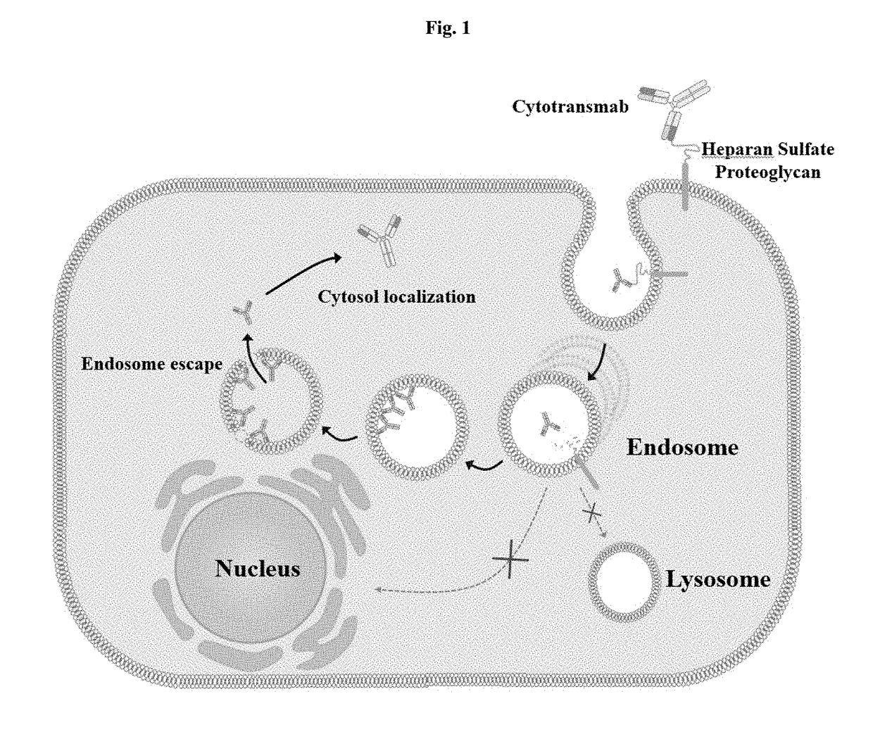
[0177]FIG. 1 is a schematic view showing the concept of an intact immunoglobulin antibody, named “cytotransmab”, which penetrates a cell and localizes in the cytosol. To realize this antibody and understand the cytosol-penetrating ability of humanized antibody light-chain variable regions, reference was made to conventional studies on the correlations between the cytosol-penetrating ability of the mouse light-chain variable single domain m3D8 VL and CDRs corresponding to light-chain variable region fragments (Lee et al., 2013).

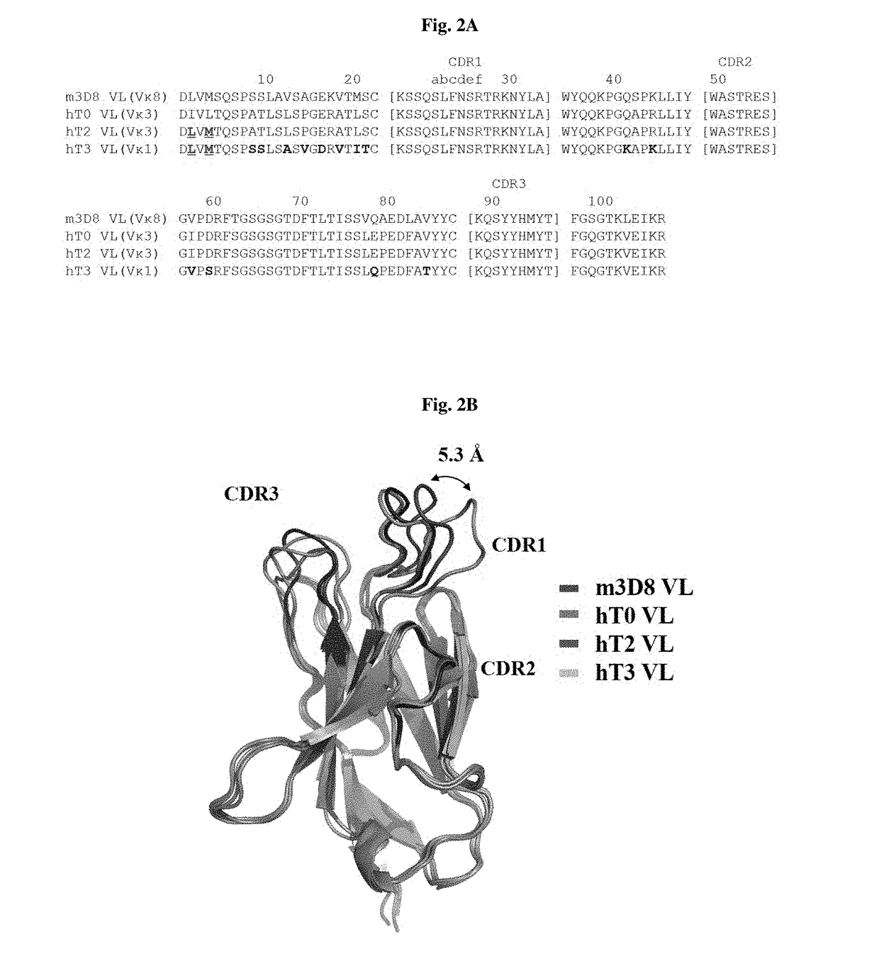
[0178]FIG. 2A shows the results of analysis of a sequence including a clone used in a process of obtaining the improved, cytosol-penetrating humanized light-chain variable single domain hT3 VL, which binds stably to a humanized antibody heavy-chain variable region, from the mouse light-chain variable region m3D8 VL.

[0179]Specifically, based on a comparison of cytosol-penetr...

example 2

n and Purification of Humanized Light-Chain Variable (VL) Single Domain Having Cytosol-Penetrating Ability

[0183]To compare the actual cytosol-penetrating abilities of hT2 VL and hT3 VL designed in the above Example, humanized light-chain variable (VL) single domains were purified.

[0184]Specifically, the cytosol-penetrating light-chain variable single domain containing a Pho A signal peptide at the N-terminus and a protein A tag at the C-terminus was cloned into a pIg20 vector by NheI / BamHI restriction enzymes, and then the vector was transformed into E. coli BL21(DE3)plysE for protein expression by electroporation. The E. coli was cultured in LBA medium containing 100 ug / ml of ampicillin at 180 rpm and 37° C. until the absorbance at 600 nm reached 0.6-0.8. Then, the culture was treated with 0.5 mM of IPTG (isopropyl β-D-1-thiogalactopyronoside, and then incubated at 23° for 20 hours to express the protein. After expression, the culture was centrifuged by a high-speed centrifuge at 8...

example 3

ion of Cytosol-Penetrating Ability and Cell Penetration Mechanism of Cytosol-Penetrating Humanized Light-Chain Variable (VL) Single Domain

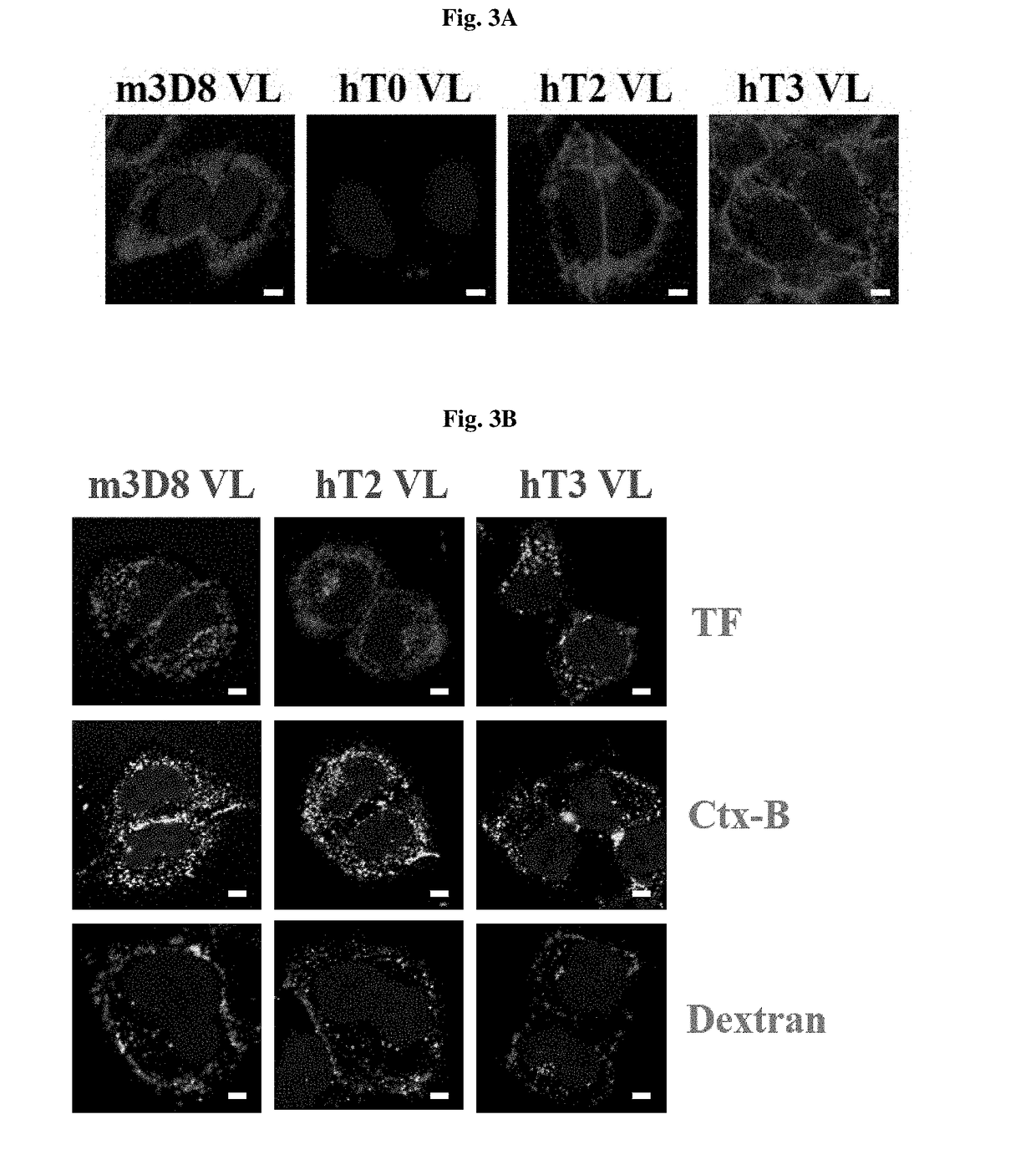
[0185]FIG. 3A shows the results of confocal microscopy observation of the cytosol-penetrating ability of light-chain variable single domains.

[0186]Specifically, in order to verify the cytosol-penetrating abilities of m3D8 VL, hT0 VL, hT2 VL and hT3 VL, a cover slip was added to 24-well plates, and 5×10⁴ HeLa cells per well were added to 0.5 ml of 10% FBS (Fetal bovine Serum)-containing medium and cultured for 12 hours under the conditions of 5% CO2 and 37° C. When the cells were stabilized, each well was treated with 10 μM of m3D8 VL, hT0 VL, hT2 VL or hT3 VL in 0.5 ml of fresh medium, and incubated for 6 hours under the conditions of 37° C. and 5% CO2. Next, the medium was removed, and each well was washed with PBS, and then treated with a weakly acidic solution (200 mM glycine, 150 mM NaCl, pH 2.5) to remove proteins from the cell surface. Next,...


PUM

PropertyMeasurementUnit
Lengthaaaaaaaaaa
Lengthaaaaaaaaaa
Lengthaaaaaaaaaa

Abstract

The present invention relates to a method of localizing an intact immunoglobulin-format antibody in cytosol by permeating membrane of cells. The present invention also relates to a light-chain variable region (VL) that induces an intact immunoglobulin-format antibody to penetrate the membrane of living cells and be localized in the cytosol, and to an antibody comprising the same. The present invention also relates to a biologically active molecule fused to the antibody and selected from the group consisting of peptides, proteins, small-molecule drugs, nanoparticles and liposomes. The present invention also relates to a composition for prevention, treatment or diagnosis of cancer, comprising: the antibody; or a biological active molecule fused to the antibody and selected from the group consisting of peptides, proteins, small-molecule drugs, nanoparticles and liposomes. The present invention also relates to a polynucleotide that encodes the light-chain variable region and the antibody. The present invention also relates to a method for producing an antibody which penetrates cells and is localized in the cytosol.According to the method of the present invention, which allows an intact immunoglobulin-format antibody to actively penetrate living cells and be localized in the cytosol, the antibody can penetrate living cells and be localized in the cytosol, without having to use a special external protein delivery system. Moreover, the use of the cytosol-penetrating light-chain variable region according to the present invention and the intact immunoglobulin-format antibody comprising the same can penetrate cells and remain in the cytosol, without affecting the high specificity and affinity of a human antibody heavy-chain variable region (VH) for antigens, and thus can be localized in the cytosol which is currently classified as a target in disease treatment based on small-molecule drugs, and at the same time, can exhibit high effects on the treatment and diagnosis of tumor and disease-related factors that show structurally complex interactions through a wide and flat surface between protein and protein. In addition, these can selectively inhibit KRas mutants, which are major drug resistance-associated factors in the use of various conventional tumor therapeutic agents, and at the same time, can be used in combination with conventional therapeutic agents to thereby exhibit effective anticancer activity.

Description

CROSS REFERENCE TO RELATED APPLICATIONS[0001]This application is a U.S. national phase under the provisions of 35 U.S.C. §371 of International Patent Application No. PCT / KR15 / 07626 filed Jul. 22, 2015, which in turn claims priority of Korean Patent Application No. 10-2014-0092673 filed Jul. 22, 2014 and Korean Patent Application No. 10-2015-0103163 filed Jul. 21, 2015. The disclosures of such international patent application and Korean priority patent applications are hereby incorporated herein by reference in their respective entireties, for all purposes.TECHNICAL FIELD[0002]The present invention relates to a method of localizing an intact immunoglobulin-format antibody in cytosol by actively permeating membrane of cells.[0003]The present invention also relates to a light-chain variable region that induces an intact immunoglobulin-format antibody to be localized in cytosol by permeating a membrane of cells, and relates to an antibody comprising the same.[0004]The present invention ...

Claims


Application Information

Patent Timeline
IPC IPC(8): C07K16/32G01N33/574C07K16/30
CPCC07K16/32C07K16/30G01N33/574C07K2317/92C07K2317/56C07K2317/24C07K2317/569C07K2317/565C07K16/46C07K16/00C07K16/2863C07K16/40A61K2039/505C07K2317/515C07K2317/73C07K2317/77C07K2317/82A61P35/00A61K39/395A61K47/50
Inventor KIM, YONG-SUNGCHOI, DONG-KISHIN, SEUNG-MINKIM, SUNG-HOON
Owner ORUM THERAPEUTICS INC
Learn More