Automatic tract extraction via atlas based adaptive connectivity-based clustering
- Summary
- Abstract
- Description
- Claims
- Application Information
AI Technical Summary
Benefits of technology
Problems solved by technology
Method used
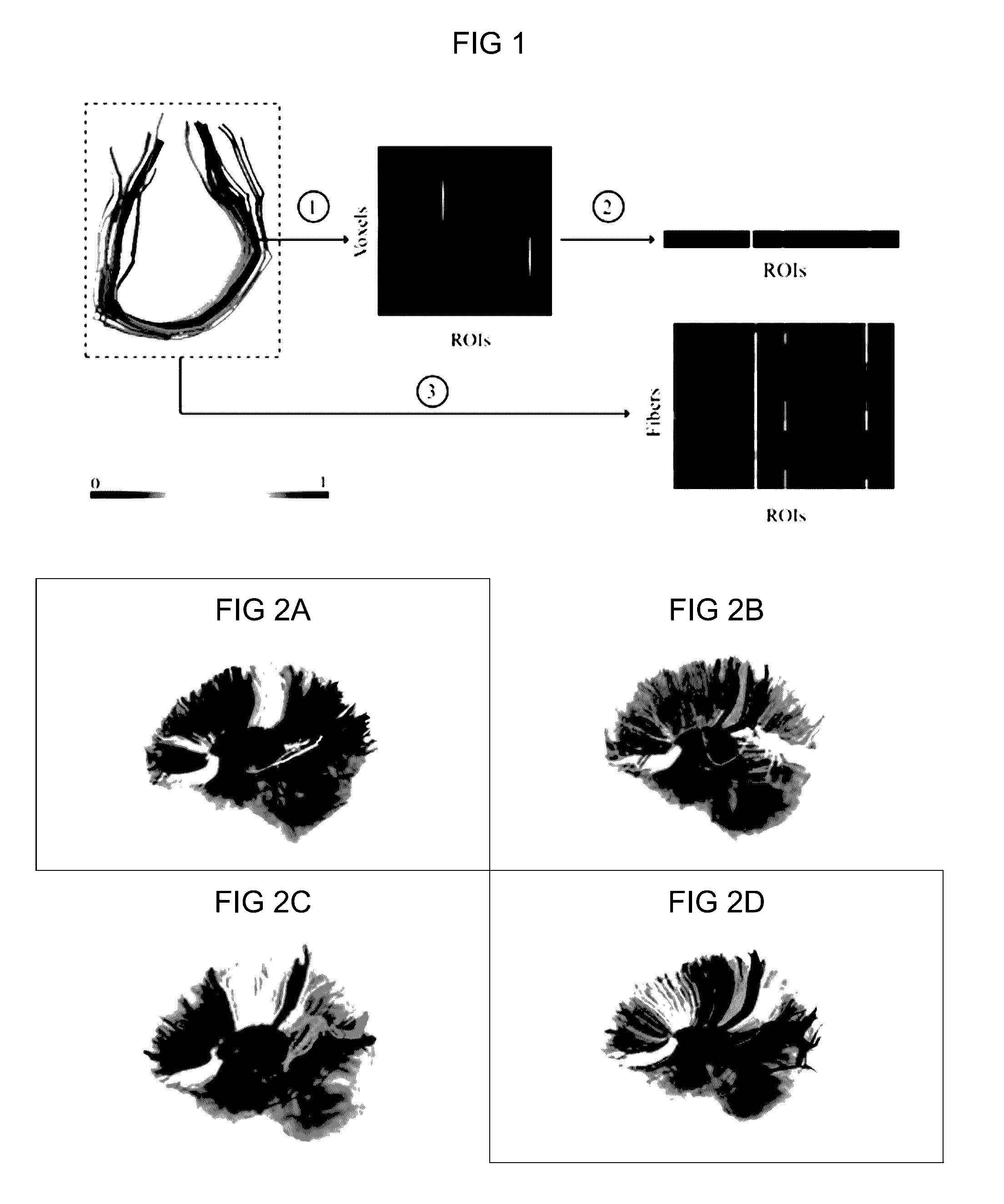
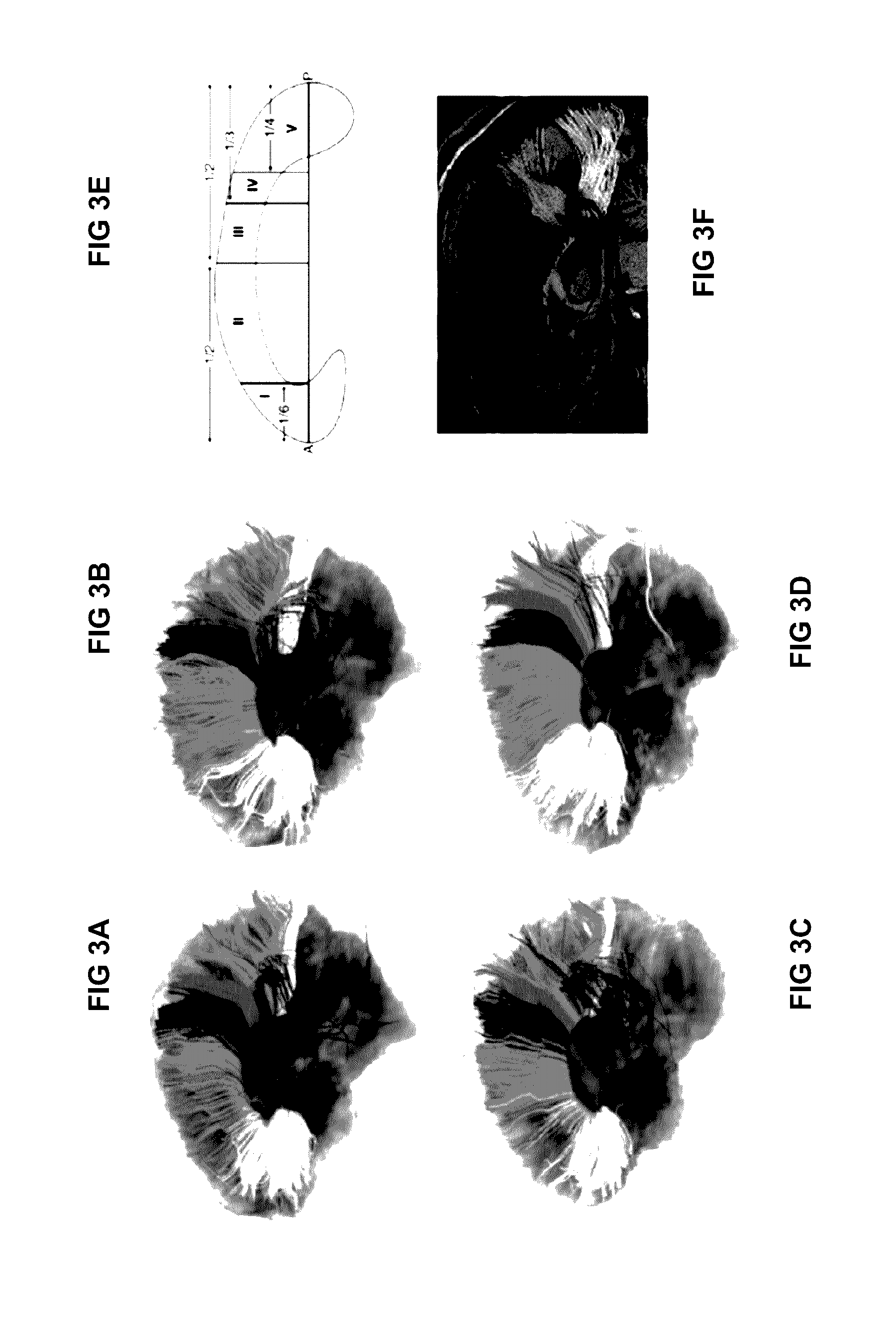
Image


Examples
example 1
[0101]The following experiments were performed to demonstrate the applicability, the reliability and the repeatability of our approach using the high angular resolution diffusion-weighted imaging (HARDI) scans of six healthy individuals each having three scans acquired at different time points. With investigations provided below, the ability of the process described herein to group and longitudinal studies to extract TOIs were validated consistently across subjects.
[0102]Dataset
[0103]Imaging was performed on six healthy male subjects (Age 31.25+4.2 years) at three time points separated by two weeks. All participants were carefully screened to ensure that they did not have a history of current or prior neuropsychiatric symptomatology. For each subject at each time point, a whole brain HARDI dataset was acquired using a Siemens 3T Verio™ scanner using a monopolar Stejskal-Tanner diffusion weighted spin-echo, echo-planar imaging sequence (TR / TE=14.8s / 111 ms, 2 mm isotropic voxels, b=30...
example 2
Automated Identification of Fiber Pathways in Patients with Edema and Tumor Mass
[0114]A. Tensor Model and Tractography
[0115]Quality assurance of the acquired data was conducted to detect artifacts and outliers, followed by diffusion weighted imaging (DWI) de-noising using Slicer [Pieper, S., et al, (2004) 3D Slicer. In: IEEE International Symposium on Biomedical Imaging; p. 632-635] and brain extraction using FSL [M. Jenkinson, et al. (2012) Neuroimage, 62(2): 782-790]. Tensors were fitted to the DWI data using multivariate linear fitting [C. Pierpaoli and Basser P J, (1996), Magn Reson Med, 36(6); 893-906] by in-house software. The WM fiber pathways were generated by the standard DTI tractography method (FACT) as implemented by TrackVis [Wang R., et al. (2007) Diffusion Toolkit: A Software Package for Diffusion Imaging Data Processing and Tractography. In Intl Soc Mag Reson Med; 3720], with default parameters and by seeding from the entire WM region. In order to calculate the conne...
example 3
Automated Identification of Fiber Pathways: Novel Paradigm for Neurosurgical Planning and Resection of Gliomas
[0120]An automated tract identification paradigm was developed and assessed for reliability in the resection of human gliomas and general neurosurgical use. A fiber bundle atlas was generated from six healthy participants. Fibers of a test set (including three healthy participants and ten patients with brain tumors) were clustered adaptively using this atlas. Reliability of identified tracts in both groups was assessed by comparison with two experts, using Cohen's kappa to quantify concurrence.
[0121]The automated paradigm demonstrated a reliable and practical method to identify white mater tracts, even when in the presence of mass effect, edema, and tract infiltration. When the tumor demonstrated significant mass effect or shift, the automated approach was useful to provide an initialization to guide the expert to identify the specific tract of interest.
[0122]Thus, this stud...
PUM
 Abstract
Description
Claims
Application Information
 - R&D
- Intellectual Property
- Life Sciences
- Materials
- Tech Scout
- Unparalleled Data Quality
- Higher Quality Content
- 60% Fewer Hallucinations
Browse by: Latest US Patents, China's latest patents, Technical Efficacy Thesaurus, Application Domain, Technology Topic, Popular Technical Reports.
© 2025 PatSnap. All rights reserved.Legal|Privacy policy|Modern Slavery Act Transparency Statement|Sitemap|About US| Contact US: help@patsnap.com