
Fast Prospective Motion Correction For MR Imaging

a prospective motion correction and imaging technology, applied in the direction of magnetic measurement, instruments, measurements using nmr, etc., can solve the problems of inability to diagnose images, mr imaging procedures are susceptible to motion of subjects, and imply a substantial cost in tim

Inactive Publication Date: 2017-01-19
SIEMENS HEALTHCARE GMBH +1
View PDF4 Cites 30 Cited by
  • Summary
  • Abstract
  • Description
  • Claims
  • Application Information

AI Technical Summary

Benefits of technology

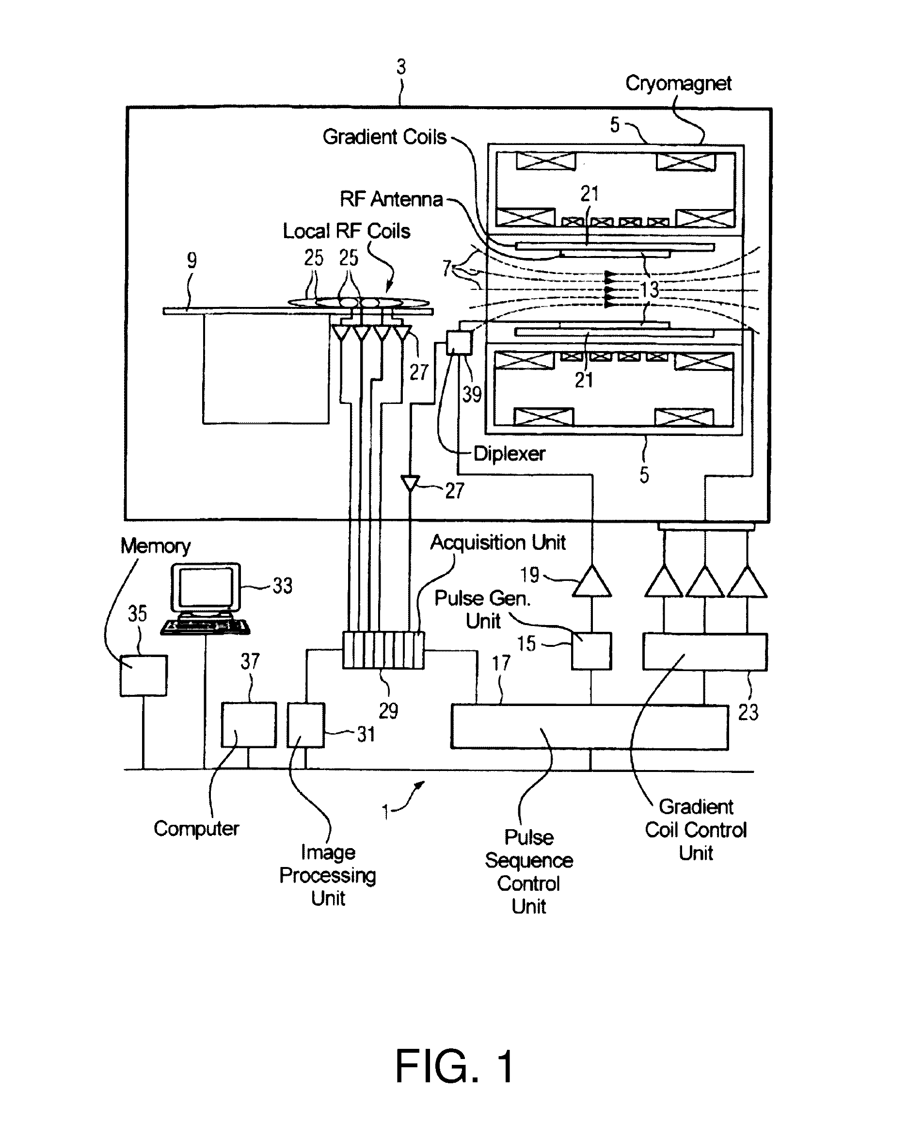
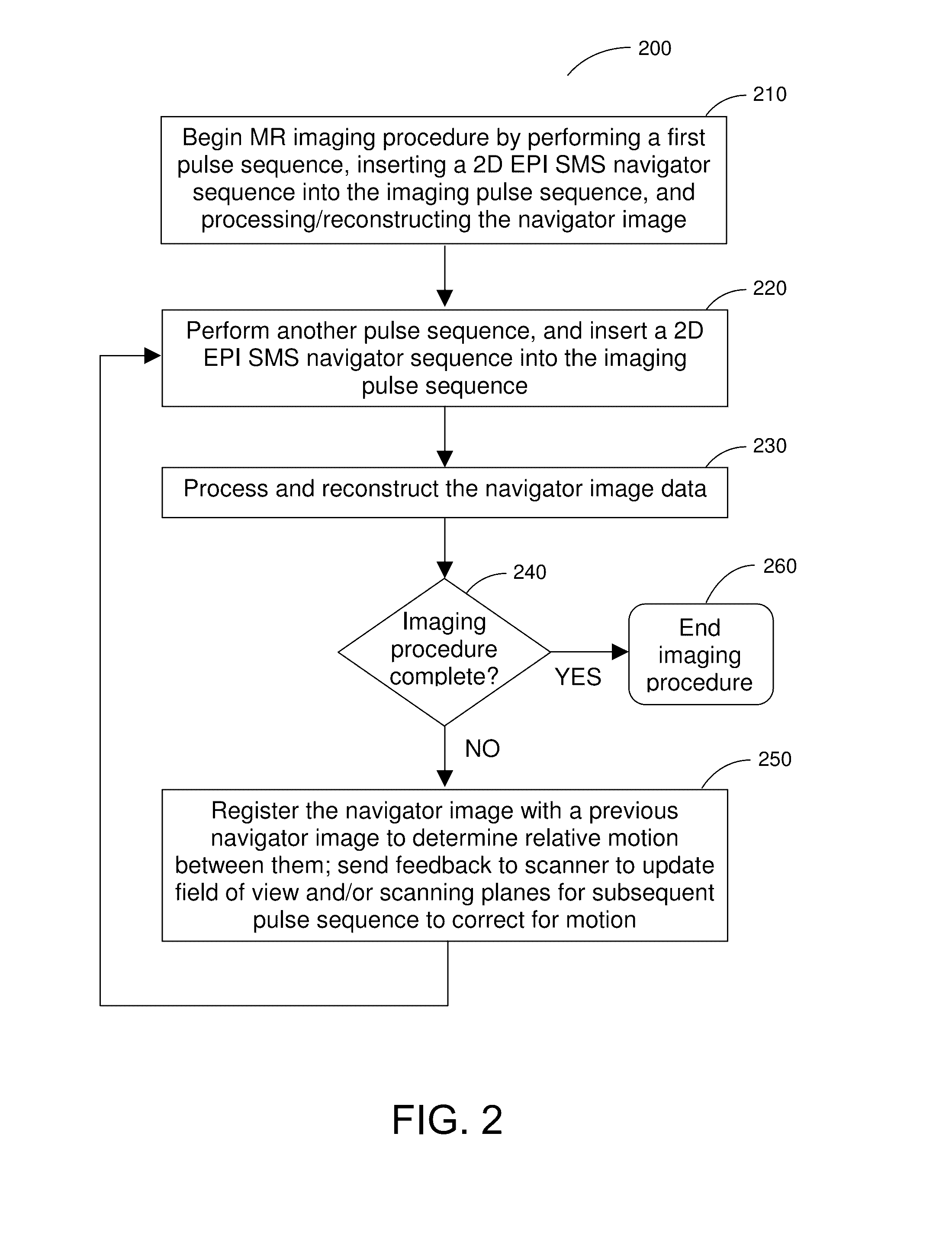
The present disclosure describes a magnetic resonance system and method for generating real-time motion-corrected images using fast navigators. The system uses a 2D EPI navigator obtained through a simultaneous multi-slice blipped-CAIPI technique to determine motion of a subject during an imaging procedure. The navigator images are obtained and processed to adapt imaging parameters for a subsequent pulse sequence, resulting in improved image quality and faster imaging with reduced motion blur. The system can be used with a wide range of MR imaging techniques, particularly those where the pulse sequences do not have significant intervals of "dead" time.

Problems solved by technology

However, such MR imaging procedures can be susceptible to motion of the subject during acquisition of imaging data.
Even a few millimeters of movement during an MRI procedure can result in motion artifacts that can render the resulting images diagnostically unusable.
One solution to the problem of motion-based image corruption is to discard and reacquire the entire scan, but this implies a substantial cost in time.
Alternatively, subject sedation can be used with certain populations (e.g., pediatric clinical subjects) but imposes a risk and is not normally considered ethical for research purposes.
One of the limitations of the EPI navigator approach is the additional time required to collect the navigator images, which typically ranges from about 170 to 300 msec, depending on spatial resolution and coverage.
This makes it difficult to insert such navigator sequences into many imaging pulse sequences that do not include significant “dead” time (e.g. 3D fast low angle shot, or FLASH, imaging pulse sequences).
Also, EPI navigators can be susceptible to motion during navigator acquisition, such that longer navigator sequences can limit their accuracy and usefulness.
However, such marker-based approaches can require additional and / or specialized equipment, and may be incompatible with certain imaging techniques and / or be less accurate because the markers can be located at some distance from the volume of interest being imaged.

Method used


Image

  • Fast Prospective Motion Correction For MR Imaging
  • Fast Prospective Motion Correction For MR Imaging
  • Fast Prospective Motion Correction For MR Imaging

Examples


example 1

[0065]To demonstrate the accuracy of the prospective motion correction system and method described herein, 2D SMS EPI navigator images were acquired of two human subjects. Each subject was asked to move their head to twelve distinct positions and orientations (relative to a particular initial location) during the imaging procedure. The images were obtained using a 3T MAGNETOM® Skyra scanner (Siemens Healthcare GmbH, Erlangen, Germany). The following parameters were used to image these navigators: FOV: 256×256×80 mm3; spatial resolution: 8×8×8 mm3 (for a voxel matrix size of 32×32×10, e.g., 10 slices of 8 mm thickness); a flip angle of 10°; two-shot SMS with 5 slices excited simultaneously per shot (SMS-5); and a relative shift between slices of FOV / 4 for the blipped-CAIPI technique.

[0066]The acquisition time for each slice group consisting of 5 slices was 14 msec, leading to a total acquisition time of 28 ms for the entire navigator volume (two shots of 5 slices each).

[0067]For comp...

example 2

[0070]To provide an indication of the range of motion identifiable with the prospective motion correction system and method described herein during MR imaging, five healthy volunteers were scanned with a 2.5-minute long SMS navigator sequence during which the five subjects were instructed to freely move their heads. The navigator parameters were the same for those used in Example 1 above, i.e.: navigator image data was obtained using a 3T MAGNETOM® Skyra scanner (Siemens Healthcare GmbH, Erlangen, Germany); FOV: 256×256×80 mm3; spatial resolution: 8×8×8 mm3 (voxel matrix size of 32×32×10, with 10 slices of 8 mm thickness); a flip angle of 10°; two-shot SMS with 5 slices excited simultaneously per shot (SMS5); and a relative shift between slices of FOV / 4 for the blipped-CAIPI technique.

[0071]The navigator images were retrospectively registered to derive a range of motion estimates within which SMS navigators are expected to function accurately. The range of motion derived from the na...

example 3

[0073]To demonstrate the efficacy of the prospective motion correction system and method described herein for an MR imaging procedure, two subjects were imaged with a conventional magnetization-prepared rapid acquisition gradient echo (MPRAGE) sequence using a 3T MAGNETOM® Skyra scanner (Siemens Healthcare GmbH, Erlangen, Germany) with a 32 channel receive coil. Parameters for the MPRAGE scan were: spatial resolution: 1×1×1 mm3; GRAPPA acceleration factor: 3; total scan time: 4 minutes.

[0074]2D SMS EPI navigators in accordance with embodiments of the present disclosure were inserted into the TI gaps of the MPRAGE sequences. The following parameters were used for the navigators obtained during the MPRAGE sequences: FOV: 256×256×80 mm3; spatial resolution: 8×8×8 mm3 (voxel matrix size of 32×32×10); a flip angle of 10°; two-shot SMS with 5 slices excited simultaneously per shot (SMS-5); and a relative shift between slices of FOV / 4 for the blipped-CAIPI technique.

[0075]During each TR a ...


PUM


Abstract

A magnetic resonance (MR) method and system are provided for generating real-time prospective motion-corrected images using fast navigators. The real-time motion correction is achieved by using a 2D EPI navigator that is obtained using a simultaneous multi-slice blipped-CAIPI technique. The navigator parameters such as field of view, voxel size, and matrix size can be selected to facilitate fast acquisition while providing information sufficient to detect rotational motions on the order of several degrees or more and translational motions on the order of several millimeters or more. The total time interval for obtaining and reconstructing navigator data, registering the navigator image, and providing feedback to correct for detected motion, can be on the order of about 100 ms or less. This prospective motion correction can be used with a wide range of MR imaging techniques where the pulse sequences do not have significant intervals of “dead” time.

Description

STATEMENT REGARDING FEDERALLY SPONSORED RESEARCH[0001]The present invention was made with government support under Grants R21AG046657 and ROOEB012107 awarded by NIH. The government has certain rights in the invention.FIELD OF THE DISCLOSURE[0002]The present disclosure relates to a method and a system for generating magnetic resonance (MR) images, and in particular to a method and a system for generating magnetic resonance images with fast prospective motion correction that can be used with a variety of MR imaging techniques.BACKGROUND INFORMATION[0003]Magnetic resonance (MR) imaging is a known technology that can produce images of the inside of an examination subject without radiation exposure. In a typical MR imaging procedure, the subject is positioned in a strong, static, homogeneous base magnetic field BO (having a field strength that is typically between about 0.5 Tesla and 3 Tesla) in an MR apparatus, so that the subject's nuclear spins become oriented along the base magnetic ...

Claims


Application Information

Patent Timeline
Patent Type & Authority Applications(United States)
IPC IPC(8): G01R33/565G01R33/561
CPCG01R33/56509G01R33/5611G01R33/5616G01R33/54G01R33/5602G01R33/5617G01R33/56341G01R33/56366G01R33/5676
Inventor BHAT, HIMANSHUHEBERLEIN, KEITH AARONCAULEY, STEPHEN FARMANTISDALL, MATTHEW DYLANSETSOMPOP, KAWINVAN DER KOUWE, ANDRE JAN WILLEM
Owner SIEMENS HEALTHCARE GMBH
Learn More