
Device for treating ocular diseases caused by increased intraocular pressure

Inactive Publication Date: 2016-10-20
SAMSUNG LIFE PUBLIC WELFARE FOUND
View PDF4 Cites 9 Cited by
  • Summary
  • Abstract
  • Description
  • Claims
  • Application Information

AI Technical Summary

Benefits of technology

The patent text describes a device for treating ocular diseases caused by increased intraocular pressure. The device does not require a covering tube, which reduces the risk of complications. The device has a thin tube that can be easily inserted and removed. Additionally, it allows for adjustable intraocular pressure through the use of a thread and can lower pressure when needed. This device provides a safer, less invasive, and more effective treatment for glaucoma.

Problems solved by technology

A glaucoma filtration surgery for forming a detour or a fistula to drain the aqueous humor may fail to adjust the intraocular pressure because the detour is closed after the surgery and thus the aqueous humor drainage is reduced.
Depending on the type of glaucoma, that is, for refractory glaucoma such as neovascular glaucoma or secondary glaucoma associated with uveitis, the detour closure frequently occurs after the glaucoma filtration surgery, and thus the result of the surgery is not good.
Furthermore, when the tube that is installed under the conjunctiva is so thick that the tube may protrude toward an ocular surface, the tube may be exposed after a long time, resulting in dangerous situations.

Method used


Image

  • Device for treating ocular diseases caused by increased intraocular pressure
  • Device for treating ocular diseases caused by increased intraocular pressure
  • Device for treating ocular diseases caused by increased intraocular pressure

Examples


embodiment 1

of Glaucoma Treatment Device and Surgery Using the Same

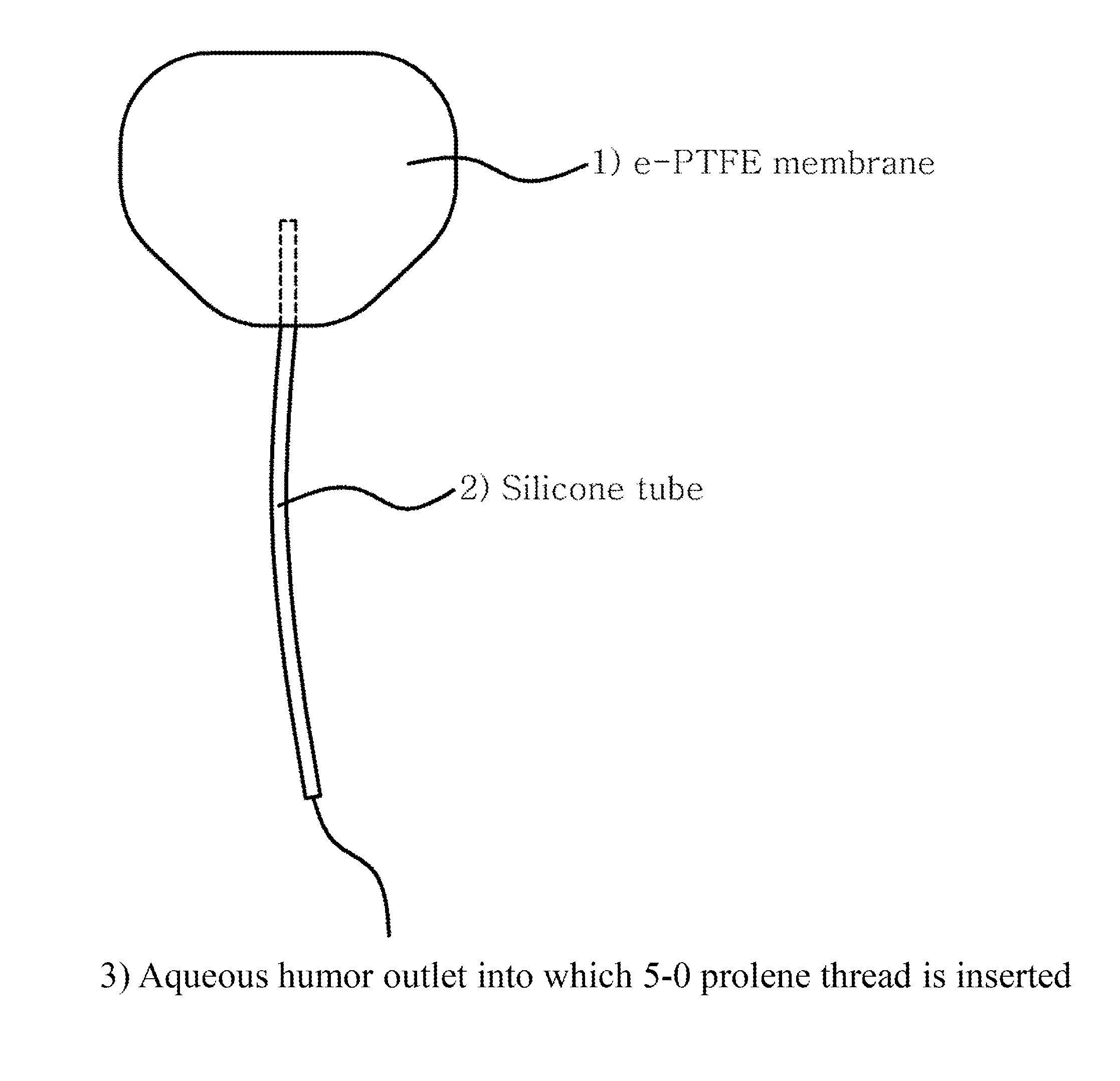
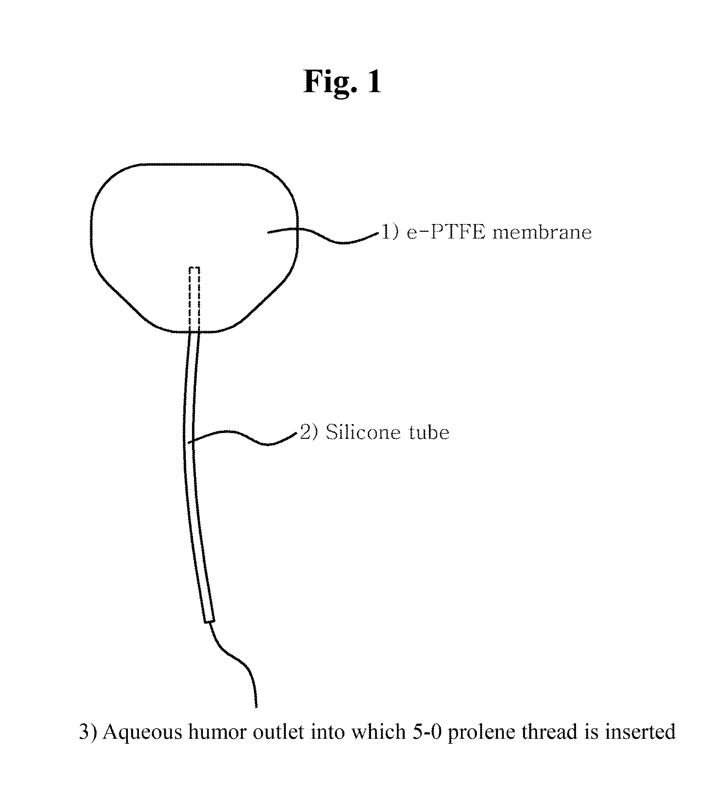
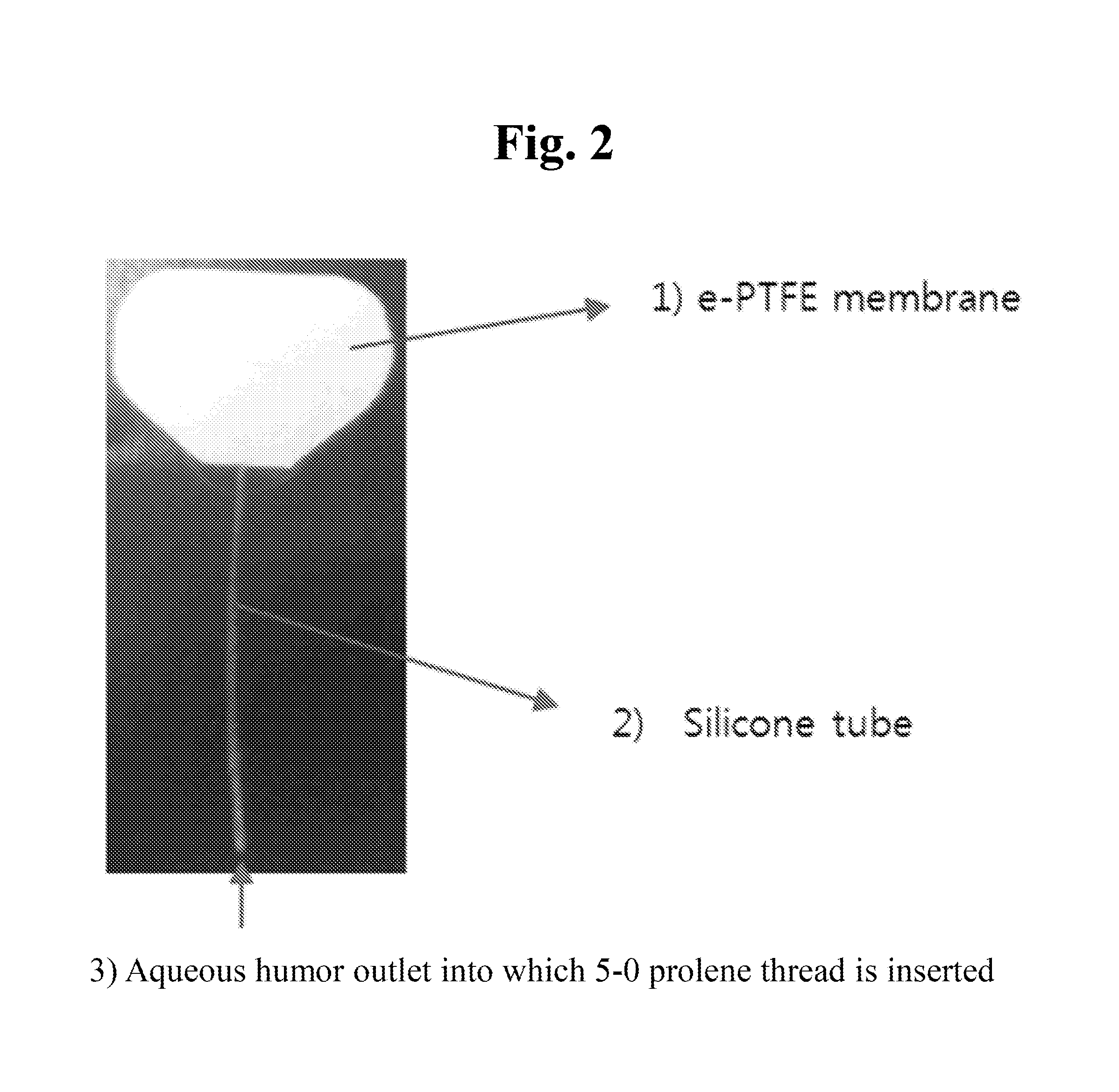
[0049]First, an expanded polytetrafluoroethylene (e-PTFE) membrane was cut to a width of about 18 mm and a length of about 13 mm, and was shaped into an oval with an area of 180 mm2 Next, a silicone tube with an internal diameter of 200 μm and an external diameter of 300 μm was inserted between double layers of the oval e-PTFE membrane and was fixed with silicone adhesive. Then the membrane was sealed at the edge with the silicone adhesive while a portion of the edge corresponding to an opposite side of the tube was not sealed so that drained aqueous humor might flow out. A 5-0 prolene thread was inserted into the fixed silicone tube, and then was pushed into an inner space of the membrane. This is to block the inside of the tube in order to prevent early postoperative hypotony.

[0050]In the department of ophthalmology at Samsung Medical Center, glaucoma surgery was performed on 30 glaucoma patients using the glaucoma treatment d...

experimental example 1

One-Year Observation After Surgery Using Glaucoma Treatment Device

[0051]Clinical outcomes were compared for patients on whom a one-year observation after the surgery using a fine tube (that is, the glaucoma treatment device) has been performed, by using the same items as those of ABC (Ahmed Baerveldt Comparison) research result (Donald L. et al, Treatment Outcomes in the Ahmed Baerveldt Comparison Study after 1 Year of 20 Follow-up. Ophthalmology. 2011; 118:443-452), which was obtained through comparison in one-year prognosis between Ahmed valve implant and Baerveldt implant. The results were summarized in Table 1 below.

TABLE 1Comparative example 1Comparative example 2Embodiment 1—(Ahmed valve group)(Baerveldt implant group)(Fine tube group)Baseline———IOP (mmHg)31.2 ± 11.231.8 ± 12.535.7 ± 12.7Glaucoma medication3.4 ± 1.13.5 ± 1.12.6 ± 0.8No. (% of baseline)143133301 day———IOP (mmHg)10.0 ± 7.9 18.6 ± 13.712.0 ± 9.6 No. (% of baseline)142(99%)130(98%)30(100%)1 week———IOP(mmHg)10.6 ± ...

experimental example 2

Comparison of Causes of Treatment Failure

[0053]The failure causes of the surgery in embodiment 1, comparative example 1, and comparative example 2 were summarized in Table 2 below.

TABLE 2Comparative example 1Comparative example 2Embodiment 1—(Ahmed valve group)(Baerveldt implant group)(Fine tube group)Adjustment of abnormal9(6%)6(5%)0IOP without additionalglaucoma surgeryGlaucoma revision surgery11(8%)1(1%)2(7%)Explantation for complication1(1%)3(2%)0(0%)Persistent hypotony0(0%)2(2%)0(0%)No light perception2(1%)6(5%)0(0%)Total23(16%)18(14%)2(7%)

[0054]As shown in Table 2, in the conventional ABC research (i.e., comparative example 1 and comparative example 2), revision surgery performed within one year from the surgery, failure in adjustment of intraocular pressure to 21 mmHg or higher regardless of using an intraocular pressure medication, explantation of the device due to complications, persistent hypotony, and no light perception were considered as the treatment failures. In embod...


PUM


Abstract

One or more example embodiments relate to a device for treating ocular diseases caused by an increased intraocular pressure. The device for treating ocular diseases according to one or more example embodiments, compared to a conventional Ahmed valve implant and Baerveldt implant, does not require a donated tissue, reduces tube exposure danger, allows intraocular pressure to be easily controlled after surgery, has a slight effusion of aqueous humor to the surrounding due to a small perforated window, decreases the types and number of postoperative interventions, and decreases the number of patients with early postoperative complications.

Description

TECHNICAL FIELD[0001]This application claims the benefit of Korean Patent Application No. 10-2013-0114037, filed on Sep. 25, 2013, in the Korean Intellectual Property Office, the disclosure of which is incorporated herein in its entirety by reference.[0002]One or more example embodiments relate to a device for treating ocular diseases caused by an increased intraocular pressure.BACKGROUND ART[0003]Intraocular pressure of glaucoma patients that cannot be adjusted by an intraocular pressure lowering agent is lowered by forming a detour for allowing aqueous humor in an anterior chamber of an eye to drain way under a conjunctiva. A glaucoma filtration surgery for forming a detour or a fistula to drain the aqueous humor may fail to adjust the intraocular pressure because the detour is closed after the surgery and thus the aqueous humor drainage is reduced. When the glaucoma filtration surgery should be performed due to the failure of the first surgery, the frequency of closure of the det...

Claims


Application Information

Patent Timeline
IPC IPC(8): A61F9/007A61M27/00
CPCA61F9/00781A61M2210/0612A61M27/006
Inventor AHN, BYUNG HEON
Owner SAMSUNG LIFE PUBLIC WELFARE FOUND
Learn More