Temperature monitoring apparatus and method for monitoring a temperature within a tissue

a temperature monitoring apparatus and tissue technology, applied in the field of temperature monitoring apparatus for monitoring a temperature within a tissue, can solve the problems of high recurrence rate, inability to monitor and control the size of ablation, and high cost of technique, and achieve the effect of low technical effort and high precision

Inactive Publication Date: 2016-08-25
KONINKLJIJKE PHILIPS NV
View PDF3 Cites 1 Cited by
  • Summary
  • Abstract
  • Description
  • Claims
  • Application Information

AI Technical Summary

Benefits of technology

[0015]It is therefore an object of the present invention to provide an improved temperature monitoring apparatus and an improved temperature monitoring method for monitoring a temperature within a tissue having a high precision with low technical effort.

Problems solved by technology

One common reason for the high recurrence rate is the inability to monitor and to control the ablation size of the thermal ablation technique to adequately destroy all tumor cells.
A temperature profile of the surgery can currently be achieved by magnet resonance-based temperature imaging, however, this technique is expensive and may not be readily available.
Alternatively ultrasound thermometry can be used for thermal ablation monitoring, however, since the sensitivity of this method is significantly reduced above 50° C. and since the ultrasound echoes significantly change with the onset of tissue necrosis, a precise temperature measurement and the generations of a precise temperature map of the ablation zone is not possible.
However, the heat introduction during radiofrequency microwave ablation forms gas bubbles around the core of the ablation zone, which can introduce artifacts in the shear wave imaging so that the thermal monitoring of the ablation can be disturbed.
Both ultrasound inspection methods using ultrasound thermometry or shear wave imaging can be disturbed by metal devices so that the monitoring close to the core of the ablation zone is not possible due to the presence of the radio frequency ablation probe.
Alternative methods dispose the shear wave imaging planes on the periphery of the ablation zone and determine the temperature of the ablation zone by using a thermal model, however, due to the inhomogeneity of the tissue due to blood vessels, which act as heat sinks, a precise determination of the temperature in the ablation zone is not possible.
US 2010 / 0256530 A1 discloses a method and an apparatus for monitoring tissue ablation, wherein the position of the ablation electrodes is monitored by an ultrasound transducer, however, due to the metal electrodes and the temperature and gas bubble effects in the ablation zone, a precise temperature monitoring of the ablation zone is not possible.

Method used


Image

  • Temperature monitoring apparatus and method for monitoring a temperature within a tissue
  • Temperature monitoring apparatus and method for monitoring a temperature within a tissue
  • Temperature monitoring apparatus and method for monitoring a temperature within a tissue

Examples


Embodiment Construction

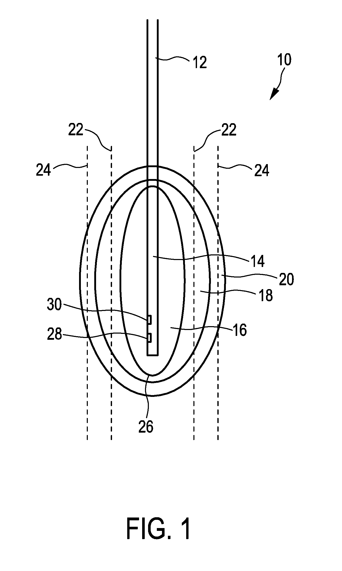
[0046]FIG. 1 shows a tissue e.g. of a human being generally denoted by 10. In the tissue 10, a temperature application device 12 having a probe or a tip 14 is disposed for heating the tissue 10 to a predefined temperature in order to coagulate parts of the tissue 10 or to ablate parts of the tissue 10. The temperature application device 12 is preferably a radio frequency application unit, a cryotherapy unit or a microwave ablation unit as minimally invasive techniques or may alternatively be a non-invasive heat source such as using ultrasound (HIFU). The temperature application device 12 introduces heating power into the tissue 10 so that the tissue 10 is heated to a temperature above 60° C. and that the tissue 10 in an ablation zone 16 is ablated or coagulated. Due to the thermal conductivity of the tissue 10, the ablation zone 16 is surrounded by medium temperature zone 18, wherein the tissue 10 in the medium temperature zone 18 is heated to a medium temperature lower than the tem...


PUM


Abstract

A temperature monitoring apparatus (40) is disclosed for monitoring a temperature within a tissue (10), in particular during a thermal ablation process. The monitoring apparatus comprises a temperature application unit (42) configured to introduce heating power into the tissue for heating the tissue. The monitoring apparatus further comprises an ultrasound unit (44) for emitting and receiving ultrasound waves and for determining a temperature in the measurement region (22, 24) of the tissue on the basis of ultrasound shear wave detection. The monitoring apparatus further comprises a temperature estimation unit (46) including a heat transfer model (48) for estimating a temperature in a region of interest (26) within the tissue, wherein the heat transfer model is based on medical images of the tissue.

Description

FIELD OF THE INVENTION[0001]The present invention relates to a temperature monitoring apparatus for monitoring a temperature within a tissue, in particular during a thermal ablation process and to a temperature monitoring method for monitoring a temperature within a tissue, in particular during a thermal ablation process.BACKGROUND OF THE INVENTION[0002]In the field of minimally invasive surgeries thermal ablation techniques are well known and provide excellent alternatives to major surgeries. This technique merely requires needles or probes for providing a radiofrequency, cryotherapy and / or microwave ablation. Alternatively a non-invasive heat source can be used such as using ultrasound. In each case, tissue is coagulated by heating the tissue to a temperature above 60° C. This technique is preferably used to treat cancer.[0003]Radio frequency ablation is currently the most common minimally invasive heating therapy to treat cancer, in particular liver cancer. The radio frequency ab...

Claims


Application Information

Patent Timeline
Patent Type & Authority Applications(United States)
IPC IPC(8): A61B18/14A61N7/02A61B18/18A61B8/08A61B18/02
CPCA61N7/02A61B5/4836A61B5/01A61B2017/00084A61B2017/00106A61B2018/00791A61B18/02A61B18/12A61B18/18A61B2018/00803A61B2034/104A61B2034/2055A61B2034/2063A61B2034/105A61B2034/2051A61B18/14A61B2090/376A61B34/10A61B2090/374A61B34/20A61B2090/378A61B8/085A61B2018/00577A61B18/1815A61B2018/00982A61B8/485A61B5/0036
Inventor ANAND, AJAYSETHURAMAN, SHRIRAMZHOU, SHIWEIXIE, HUALI, JUNBOROBERT, JEAN-LUC
Owner KONINKLJIJKE PHILIPS NV
Why Eureka
Try Eureka