
Epimorphic Regeneration and Related Hidrogel Delivery Systems

Active Publication Date: 2015-11-12
THE WISTAR INST OF ANATOMY & BIOLOGY +1
View PDF5 Cites 4 Cited by
  • Summary
  • Abstract
  • Description
  • Claims
  • Application Information

AI Technical Summary

Benefits of technology

The patent describes a composition that can be used to promote tissue regeneration. The composition contains a substance that inhibits proline hydroxylase and a molecule called HIF1a. When the composition is in contact with cells, it can induce a regenerative response, which includes enhanced tissue remodeling, de-differentiated cellular signature, increased glycolytic enzymes, increased components of inflammatory response, and increased angiogenesis. The composition can be administered systemically or locally to a subject in need. The concentration of the HIF1a-upregulating molecule can be kept at a low level to avoid toxic side effects. The composition can release the HIF1a up-regulator at a continuous, non-toxic rate for at least 1, 4, or more weeks to enhance the pace of wound healing. The level of administration can be adjusted based on the nature and site of the wound or tissue being regenerated.

Problems solved by technology

However, the effectiveness of various PHD inhibitor compounds can be compromised by low solubility under physiological conditions, inefficient routes of administration and / or untimely delivery of therapeutic dose levels.

Method used


Image

  • Epimorphic Regeneration and Related Hidrogel Delivery Systems
  • Epimorphic Regeneration and Related Hidrogel Delivery Systems
  • Epimorphic Regeneration and Related Hidrogel Delivery Systems

Examples


example 1

Animals and In-Vivo Procedures

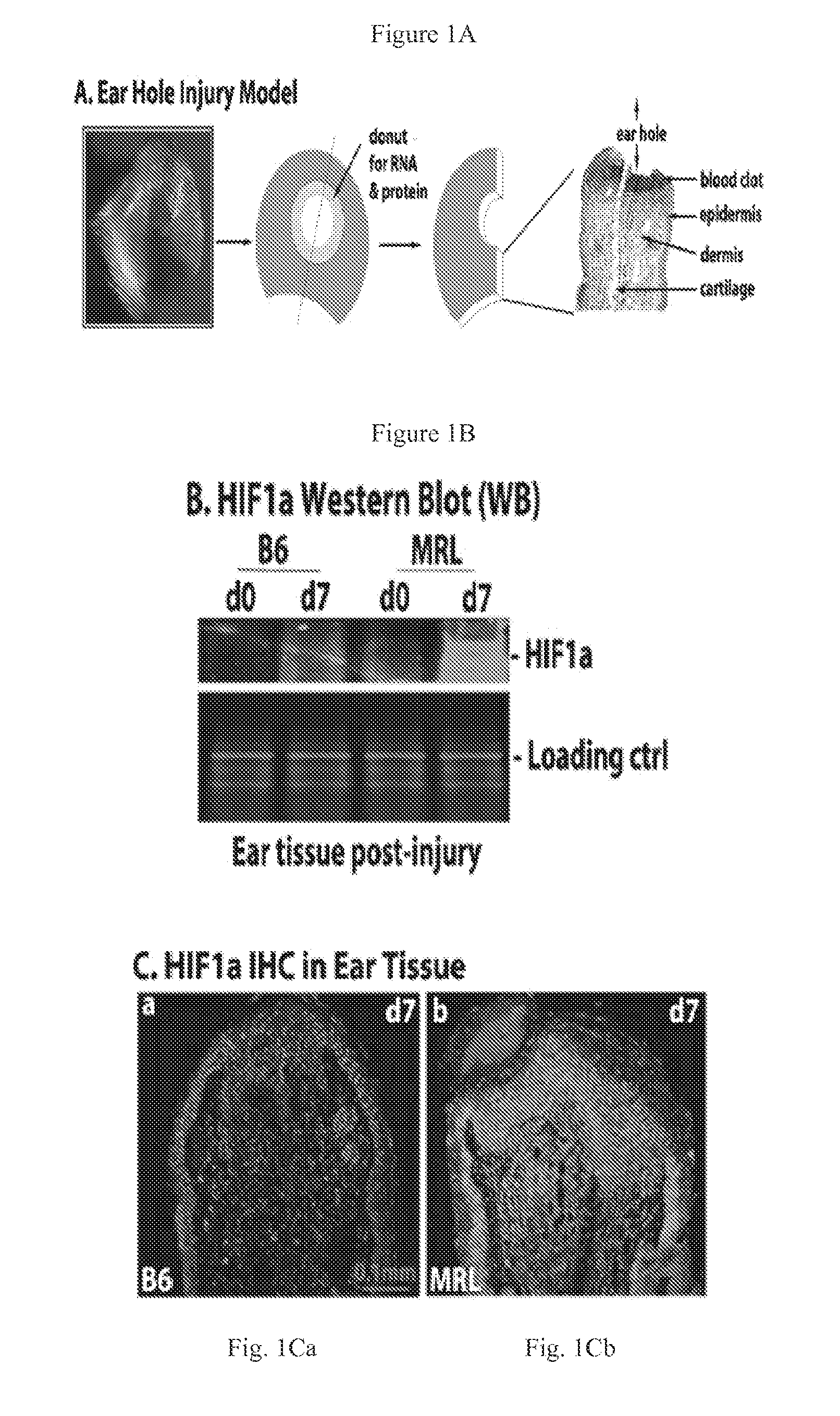
[0091]MRL / MpJ and Hif1a ODD-luciferase reporter (FVB.12956-Gt(ROSA)26Sortm2(HIF1A / luc)Kael / J) mice were obtained from Jackson Laboratories (Bar Harbor, Me.); C57BL / 6 (B6) mice were from Taconic Laboratories (Germantown, N.Y.); Swiss Webster (SW) mice were from Charles River (New York, N.Y.). Mice were used at approximately 8-10 weeks in all experiments under standard conditions at the Wistar Institute Animal Facility (Philadelphia, Pa.) and the protocols were in accordance with NIH Guide for the Care and Use of Laboratory Animals. Through-and-through ear hole punches were carried out as previously described.

example 2

IVIS Luciferase Scanning

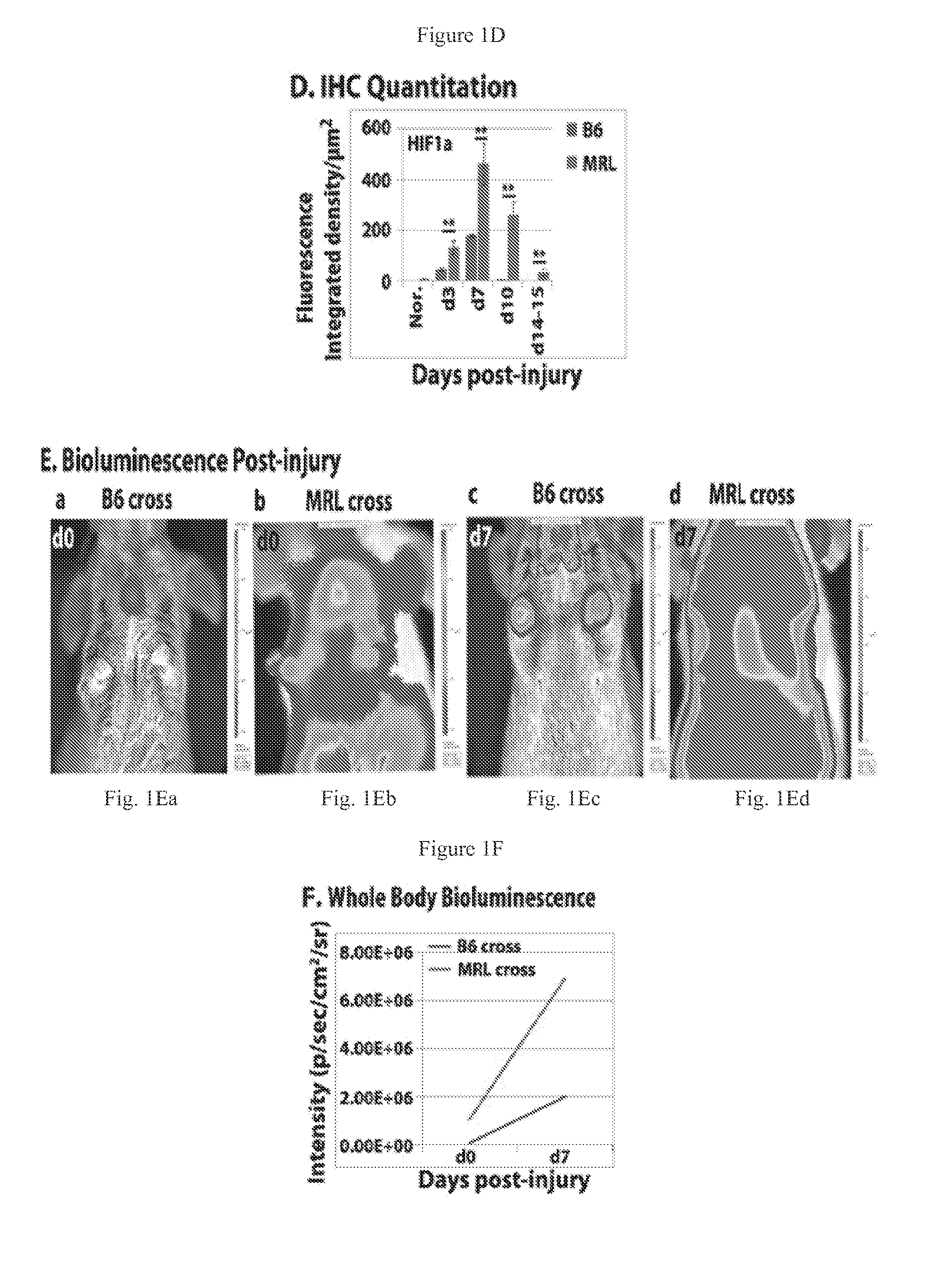
[0092]To detect luciferase expression in-vivo, mice were given a single i.p. injection of D-luciferin (37.5 mg / kg, Gold Biotechnology Inc) in sterile water. Fifteen minutes later, mice were anesthetized using isoflurane and placed in a light-tight chamber equipped with a charge-coupled device IVIS imaging camera (Xenogen, Alameda, CA). Photons were collected for a period of 1-5 min, and images were obtained by using LIVING IMAGE software (Xenogen) and IGOR image analysis software (WaveMatrics, Lake Oswego, Oreg.). HIF1a ODD luc expression after ear punching was determined in MRL and B6 mice backcrossed to the transgenic HIF1a-peptide-luciferase reporter mouse FVB.129S6-Gt(ROSA)26S, made by fusing luciferase to the domain of HIF1a that binds to pVHL in a oxygen-dependent way (ODD peptide) mice and selected for luciferase positivity.

example 3

Tissue Culture

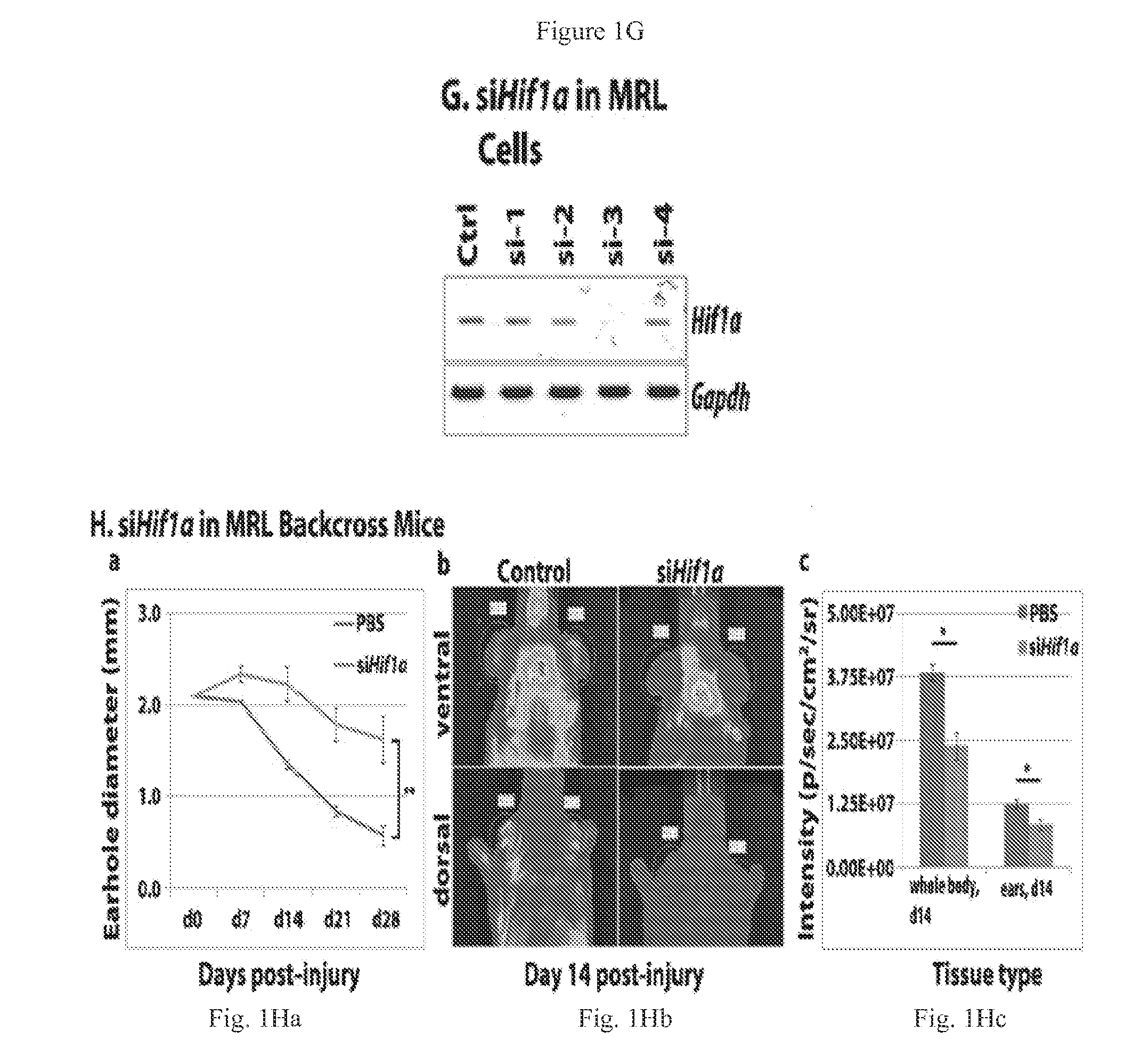
[0093]Primary ear dermal fibroblast-like cells were established from MRL and B6 mice and grown in DMEM-10% FBS supplemented with 2 mM L-glutamine, 100 IU / mL penicillin streptomycin and maintained at 37° C., 5% CO2, and 21% O2. Cells were split 1:5 as needed to maintain exponential growth and avoid contact inhibition. Passage numbers were documented and cells from early passages (<P20) frozen in liquid nitrogen and used in the described experiments.


PUM

PropertyMeasurementUnit
Timeaaaaaaaaaa

Abstract

Methods and compositions are described for enhancing tissue regeneration or wound repair in a mammalian subject comprising a composition comprising (a) a proline hydroxylase inhibitor component or molecule that increases or upregulates HIF1a and (b) a carrier component comprising a hydrogel.

Description

[0001]This application claims priority to and the benefit of application Ser. No. 61 / 962,637 filed Nov. 13, 2013, the entirety of which is incorporated herein by reference.[0002]This invention was made with government support under grant numbers RO1 DE021215, DE021104 and P30 CA010815 awarded by the National Institutes of Health. The government has certain rights in the invention.BACKGROUND OF THE INVENTION[0003]Wound repair and regeneration are two separable biological processes by which organisms heal wounds. As a model of regeneration in mammals, ear hole closure, as seen in rabbits, in the inbred mouse strains MRL / MpJ and LG / J and other mutant mouse strains shows multiple similarities to limb regeneration in amphibians including the replacement of cartilage and the lack of scarring. Other less well known classical regenerative phenotypes are also seen in the MRL ear injury model include rapid re-epithelialization, enhanced tissue remodeling, basement membrane breakdown, blastema...

Claims


Application Information

Patent Timeline
IPC IPC(8): A61K47/48A61K31/4745C12N9/02C08G65/333
CPCA61K47/48215C08G65/33396A61K31/4745C12Y114/11002C12N9/0071C08G2210/00A61K47/48784C08G65/333C08G65/332C08G65/33331C08G65/33337C08G65/3342C08G65/3344C08G65/3348C08G2650/58C08L2203/02C08G2650/32A61K47/60
Inventor MESSERSMITH, PHILLIP B.STREHIN, IOSSIF A.HEBER-KATZ, ELLEN
Owner THE WISTAR INST OF ANATOMY & BIOLOGY
Learn More