
Crystal structure of bifunctional transglycosylase pbp1b from e. coli and inhibitors thereof

a technology of bifunctional transglycosylase and crystal structure, which is applied in the field of crystal structure of bifunctional transglycosylase pbp1b from e. coli and inhibitors thereof, can solve the problems of cell lysis and death, serious medical problems, and resistance to detailed study, and achieve the effect of preventing the elongation of peptidoglycans

Inactive Publication Date: 2015-08-20
ACAD SINIC
View PDF3 Cites 1 Cited by
  • Summary
  • Abstract
  • Description
  • Claims
  • Application Information

AI Technical Summary

Benefits of technology

The patent text describes the discovery of a new antibiotic compound that can inhibit the activity of a bacterial enzyme called transglycosylase. The invention is based on the use of a three-dimensional structure of the enzyme in combination with computer modeling and drug design techniques. The new compound was found to have a higher potency for inhibiting the enzyme than existing drugs. The invention also includes a method for identifying potential inhibitors of transglycosylase using a co-crystal structure of the enzyme and a candidate agent. Overall, the invention provides a new tool for identifying and developing new antibiotics that can target transglycosylase.

Problems solved by technology

It can protect bacterial cells against osmotic pressure, and its disruption can lead to cell lysis and death.
Despite their importance to bacterial physiology and drug discovery, they have resisted detailed study, mainly because these large membrane proteins are difficult to purify, assay, and crystallize.
Not too long after they were introduced, resistant bacteria had emerged rapidly and caused serious medical problems.
During the years, resistance bacteria strains against two of the most important antibiotics, β-lactam (such as penicillin) and glycopeptide (such as vancomycin), have become a very serious medical problem in the treatment of bacterial infections.

Method used


Image

  • Crystal structure of bifunctional transglycosylase pbp1b from e. coli and inhibitors thereof
  • Crystal structure of bifunctional transglycosylase pbp1b from e. coli and inhibitors thereof
  • Crystal structure of bifunctional transglycosylase pbp1b from e. coli and inhibitors thereof

Examples


example 1

Cloning, Expression and Purification of PBP1b

[0143]Purified PBP1b gamma degraded readily into a slightly smaller protein. After N-terminal sequencing accompanying with molecular weight measurement by MALTI-TOF Mass spectrometry, we identified the stable region containing amino acid 58 to 804.

[0144]PBP1b[58-804] was amplified from E. coli genomic DNA and cloned into the expression vector pET15b (NovagEN; EMD Sciences, San Diego, Calif.)) at the NdeI and BamHI restriction sites. BL21(DE3) E. coli host cells were grown at 37° C. until OD600 reached 0.6, and protein expression was induced with 1 mM IPTG for 3 hr. Cell pellets were resuspended in 20 mM Tris at pH 8.0, 300 mM NaCl and broken by Microfluidizer™ (Microfluidics, Newton, Mass.). Recombinant protein with an N-terminal (His)6 tag was solubilized with 20 mM n-Dodecyl-β-D-maltoside (DDM; Anatrace, Maumee, Ohio, USA)) and purified by nickel chelation chromatography, in accordance with the manufacturer's instructions, in the presen...

example 2

Crystallization, Data Collection and Structure Determination

[0145]Crystals of PBP1b[58-804]-Moenomycin complex were co-crystallized in sitting drop at 16° C. Crystals were obtained by mixing 12 mg / ml protein containing additional 1.4 mM moenomycin with the same volume of reservoir solution containing 1.2 M sodium formate. For cryoprotection, crystals were transferred into 3 M sodium formate and flash-frozen in liquid nitrogen.

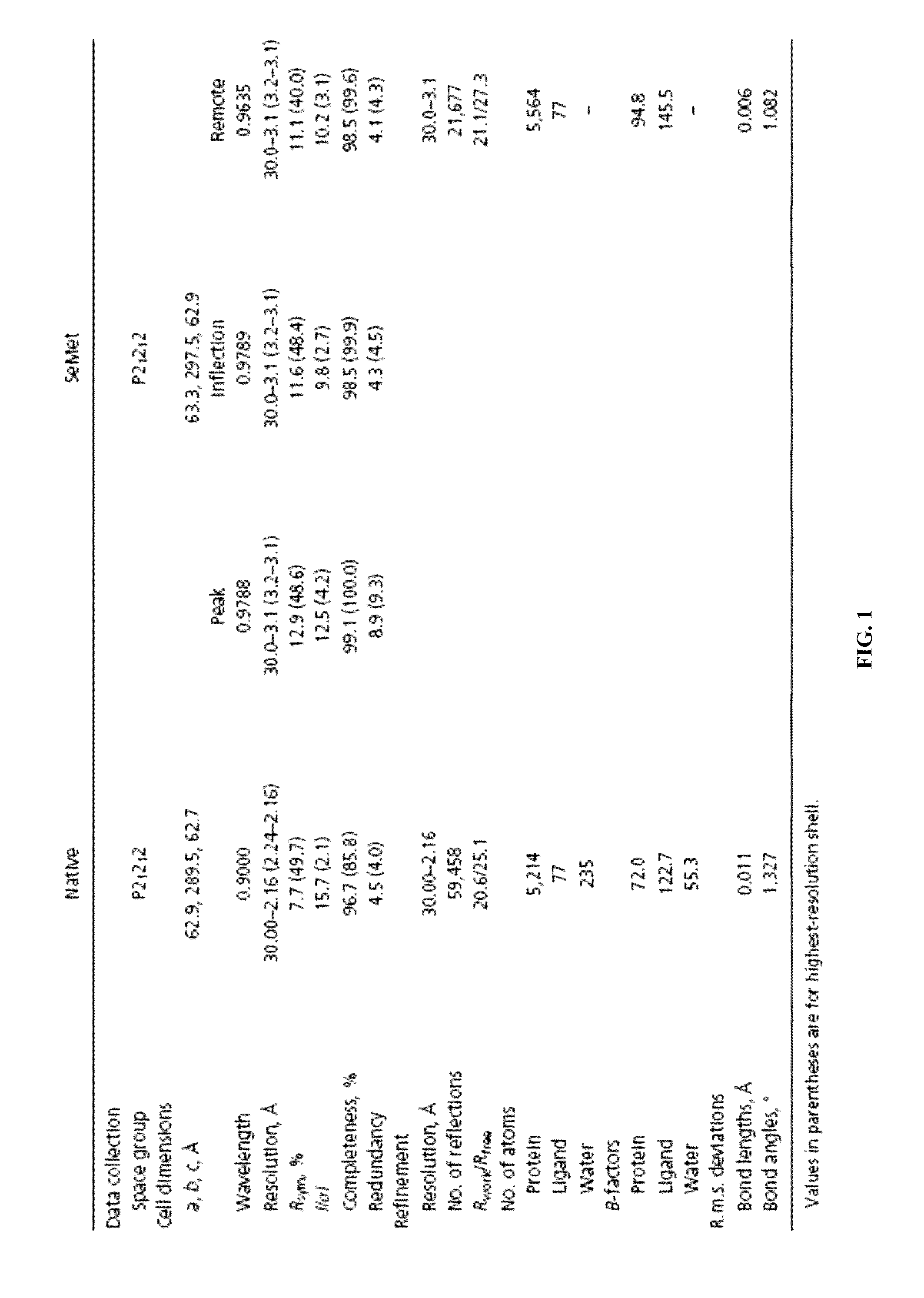
[0146]Native dataset was collected at Beamline 44XU™ of Japan Synchrotron Radiation Research Institute (Hyogo, JP) and SeMet datasets were collected at Beamline 13B1™ of National Synchrotron Radiation Research Center (Hsinchu, TW). All data were indexed, integrated and scaled with HKL2000. (Leslie, A. G., Powell, H. R., Winter, G., Svensson, O., Spruce, D., McSweeney, S., Love, D., Kinder, S., Duke, E. & Nave, C. (2002). Automation of the collection and processing of X-ray diffraction data—a generic approach. Acta Crystallogr D Biol Crystallogr 58, 1924-8.) MAD...

example 3

Molecular Dynamics Simulation

[0147]GROMACS (Van Der Spoel, D., Lindahl, E., Hess, B., Groenhof, G., Mark, A. E. & Berendsen, H. J. (2005). GROMACS: fast, flexible, and free. J Comput Chem 26, 1701-18) was used as the molecular dynamics simulation engine. MARTINI force field (Marrink, S. J. & Mark, A. E. (2004). Molecular view of hexagonal phase formation in phospholipid membranes. Biophys J 87, 3894-900) was used to model the coarse-grained PBP1b structure. After converting the atomic model into coarse-grained model, the structure was subjected to a brief steepest-descent energy minimization. It was then manually inserted into a water box containing pre-equilibrated lipid bilayer. A initial orientation of PBP1b were chosen subjectively to see if the final orientations would equilibrate to a specific position. Seven chloride ions were added into the box in order to maintain electrostatic neutrality. A steepest-descent energy minimization was carried out to relax any steric conflict b...


PUM

PropertyMeasurementUnit
temperatureaaaaaaaaaa
timeaaaaaaaaaa

Abstract

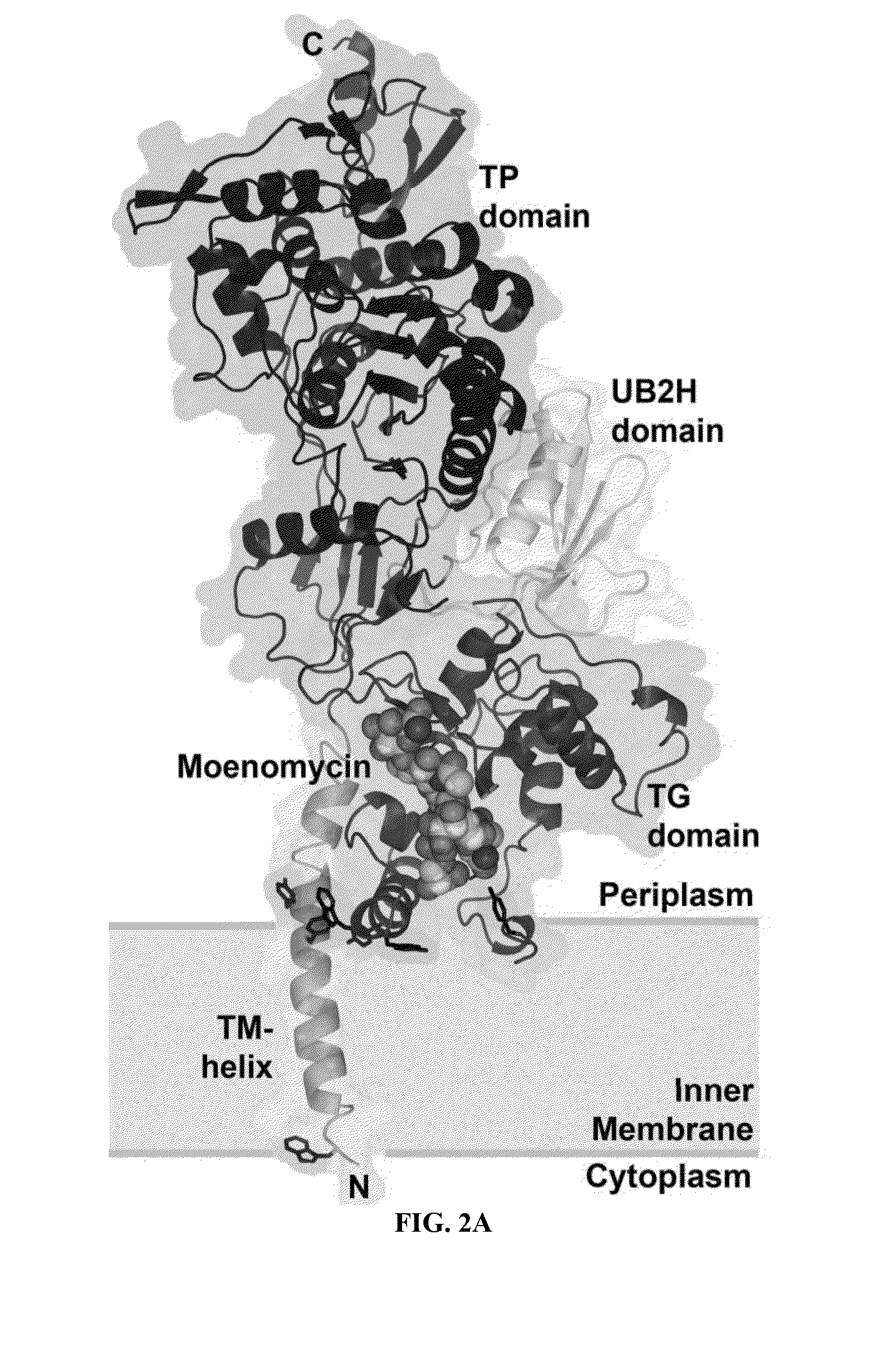
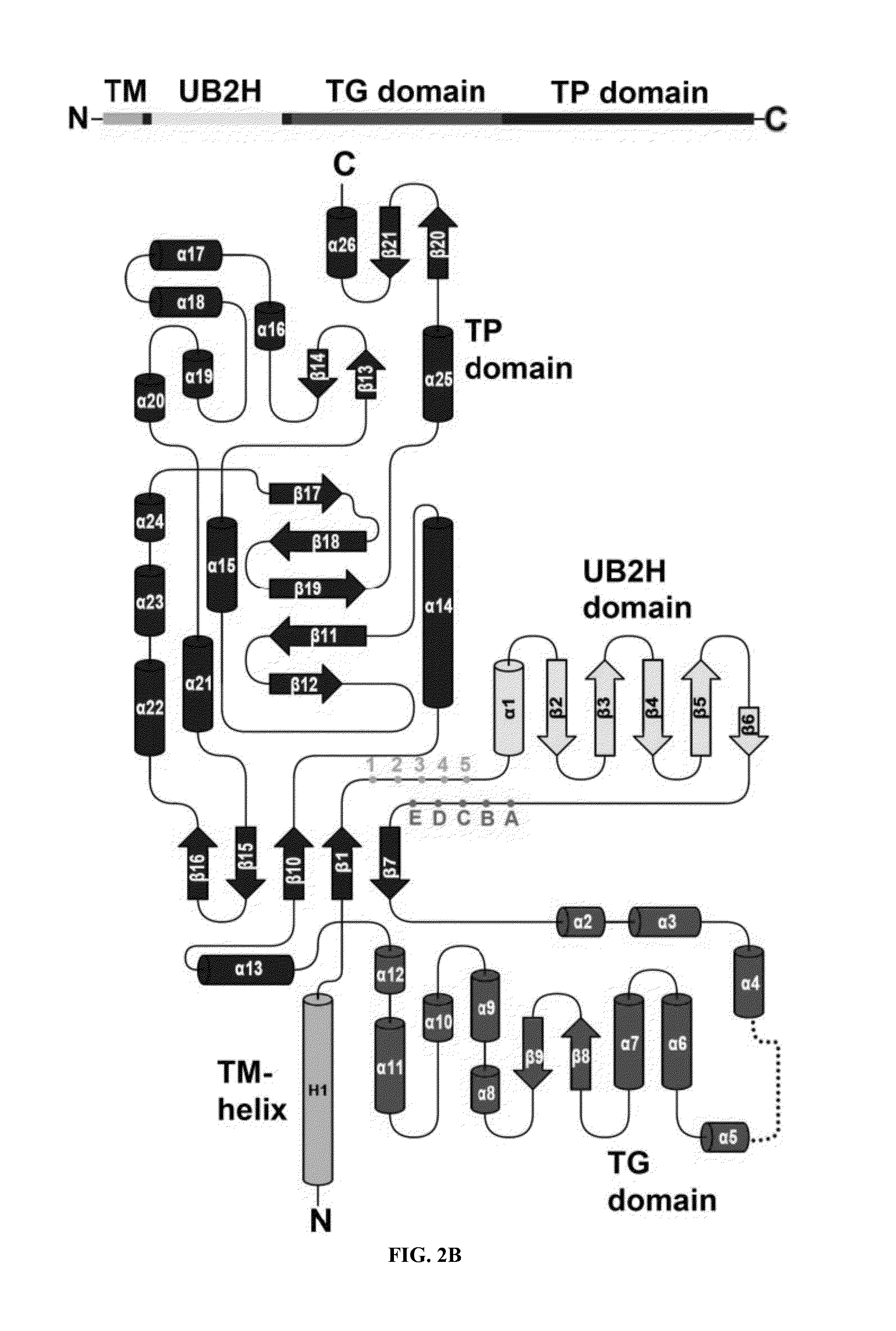
Crystal structure at 2.16 Å resolution of full-length Escherichia coli penicillin-binding protein 1b (PBP1b) in complex with its inhibitor moenomycin, is provided. 3D structures of amino acid residues involved in moenomycin binding and transglycosylation activity are identified. Binding sites for peptidoglycan synthesis inhibitors comprising amino acid residues from transglycosylase (TG), UvrB domain 2 homolog (UB2H) and transmembrane (TM) domains of PBP1b are identified at atomic level resolution. Rational drug design, based on the atomic coordinates, are disclosed. Methods for screening for antibiotics using anisotropic binding and transglycosylase inhibitor assays and novel antibiotics based on the screening assays are provided.

Description

CROSS-REFERENCE TO RELATED APPLICATIONS[0001]This application is a divisional application of U.S. Ser. No. 12 / 506,982 filed Jul. 21, 2009 which claims priority of provisional patent applications U.S. Ser. No. 61 / 208,566, titled “Structure and Functional Aspects of the Bacterial Bifunctional Transglycosylase PBP1b From E. coli And Binding Interactions With Moenomycin” filed Feb. 25, 2009, and U.S. Ser. No. 61 / 135,503, titled “Structure and Functional Aspects of the Bacterial Bifunctional Transglycosylase PBP1b From E. coli And Binding Interactions With Moenomycin” filed Jul. 21, 2008, the contents of which are incorporated herein in their entirety by reference.TECHNICAL FIELD OF THE INVENTION[0002]The present invention relates to a bacterial bifunctional Transglycosylase PBP1b from E. Coli, that can be co-crystallized with an inhibitory ligand such as moenomycin, and more particularly, to the detailed crystallographic data obtained from said co-crystallization which is disclosed here...

Claims


Application Information

Patent Timeline
Patent Type & Authority Applications(United States)
IPC IPC(8): C07C235/64G01N33/573
CPCC07K2299/00C12Q1/18C12Q1/48G01N2333/91102G01N2500/00C07C235/64C07C235/84G01N33/573G01N2500/04G01N2500/20
Inventor WONG, CHI-HUEYMA, CHE ALEXCHENG, TING-JEN RACHELCHENG, WEI-CHIEH
Owner ACAD SINIC
Learn More