Blood marker for renal cancer
a technology for blood markers and renal cancer, applied in the field of clinical diagnosis and examination, can solve the problems of not having clinically available blood markers, and the need for such markers, and achieve the effects of improving the early detection rate of renal cancer, simple and relatively cheap method, and easy detection of renal cancer
- Summary
- Abstract
- Description
- Claims
- Application Information
AI Technical Summary
Benefits of technology
Problems solved by technology
Method used
Image
Examples
example 1
Construction of ELISA System
[0063]First, ELISA measurement systems were prepared for eight kinds of proteins whose concentrations are known to increase in renal cancer tissue [α-enolase, Calnexin (for reference), CNDP dipeptidase 2 (for reference), Galectin-1, Galectin-3, Lectin mannose-binding 2 (for reference), Triosephosphate isomerase (for reference), and MHC class I antigen A (for reference)].
[0064]Among them, the measurement systems for Galectin-1 and Galectin-3 were constructed using a capture antibody, a detection antibody, and a detection reagent shown in Table 1. The detection antibody used was labeled with the labeling protein (i.e., a protein for labeling) shown in Table 1.
[0065]A capture antibody solution (5 μg / mL) was added to each of the wells of a 96-well plate (manufactured by Maxisorp) to obtain a solid-phase antibody. The solid-phase capture antibody was obtained using IMMUNO-TEK ELISA Construction System (ZeptoMetrix, Buffalo, N.Y.).
TABLE 1Galectin-1 ELISAGalecti...
example 2
[0068]Plasma samples collected from 51 healthy individuals and renal cancer patients were used to measure blood concentrations and compare the blood concentrations between the group of healthy individuals and the group of renal cancer patients. The male-to-female ratio, average age, and age range of each group of healthy individuals and renal cancer patients, the pT classification of renal cancer, presence or absence of distal metastasis, and the position of tumor (right or left kidney) are shown in Table 4.
TABLE 4Healthy individualsNumber of samples51Female (%)15(29.4)Male (%)36(70.6)Average age (Age range)62.8(40-82)Renal cancer patientsNumber of samples15Female (%)3(20.0)Male (%)12(80.0)Average age (Age range)65.1(39-79)pT classificationpT19pT22pT34Distal metastasisabsence11presence4Position of tumorLeft kidney6Right kidney9
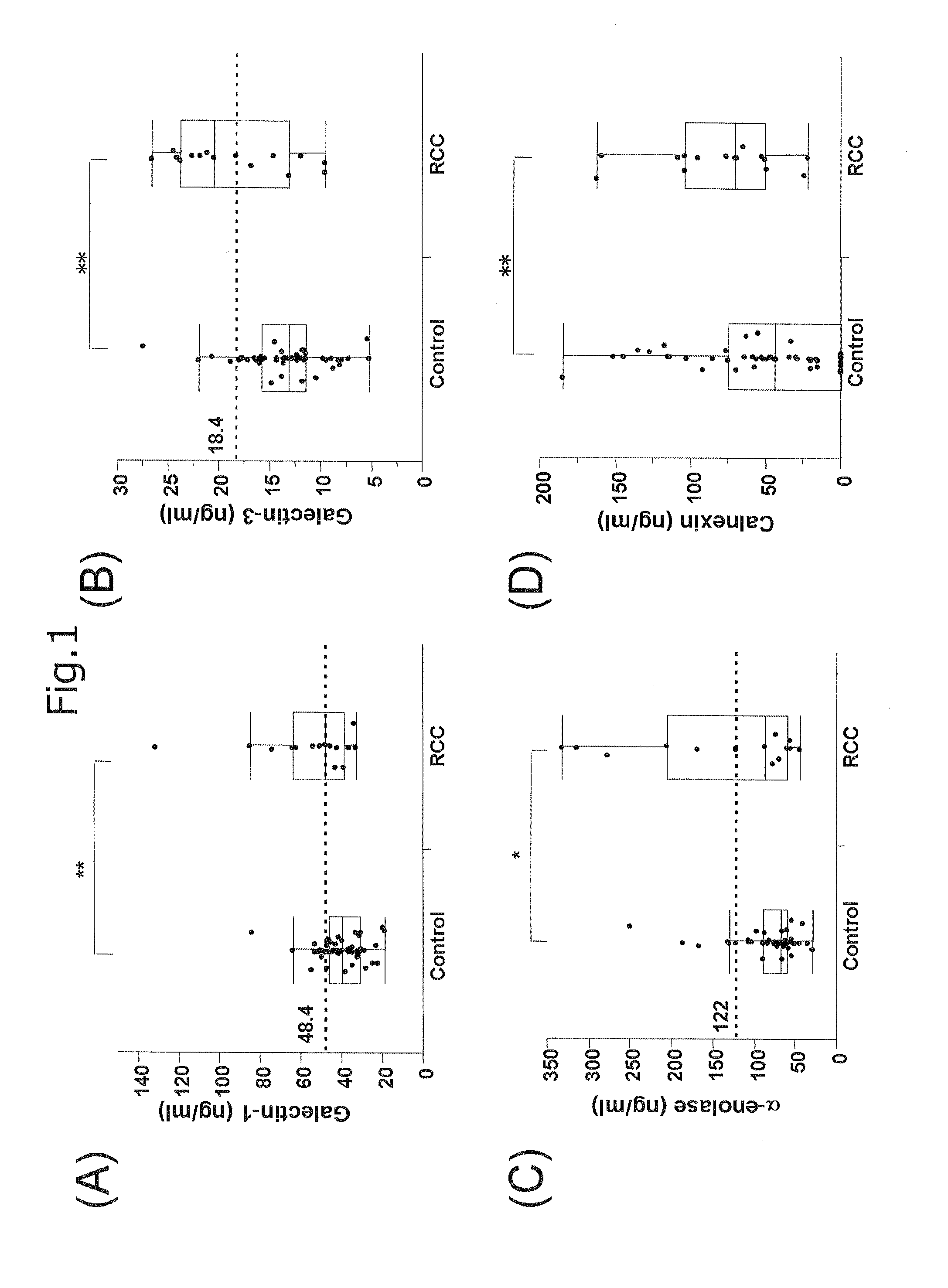
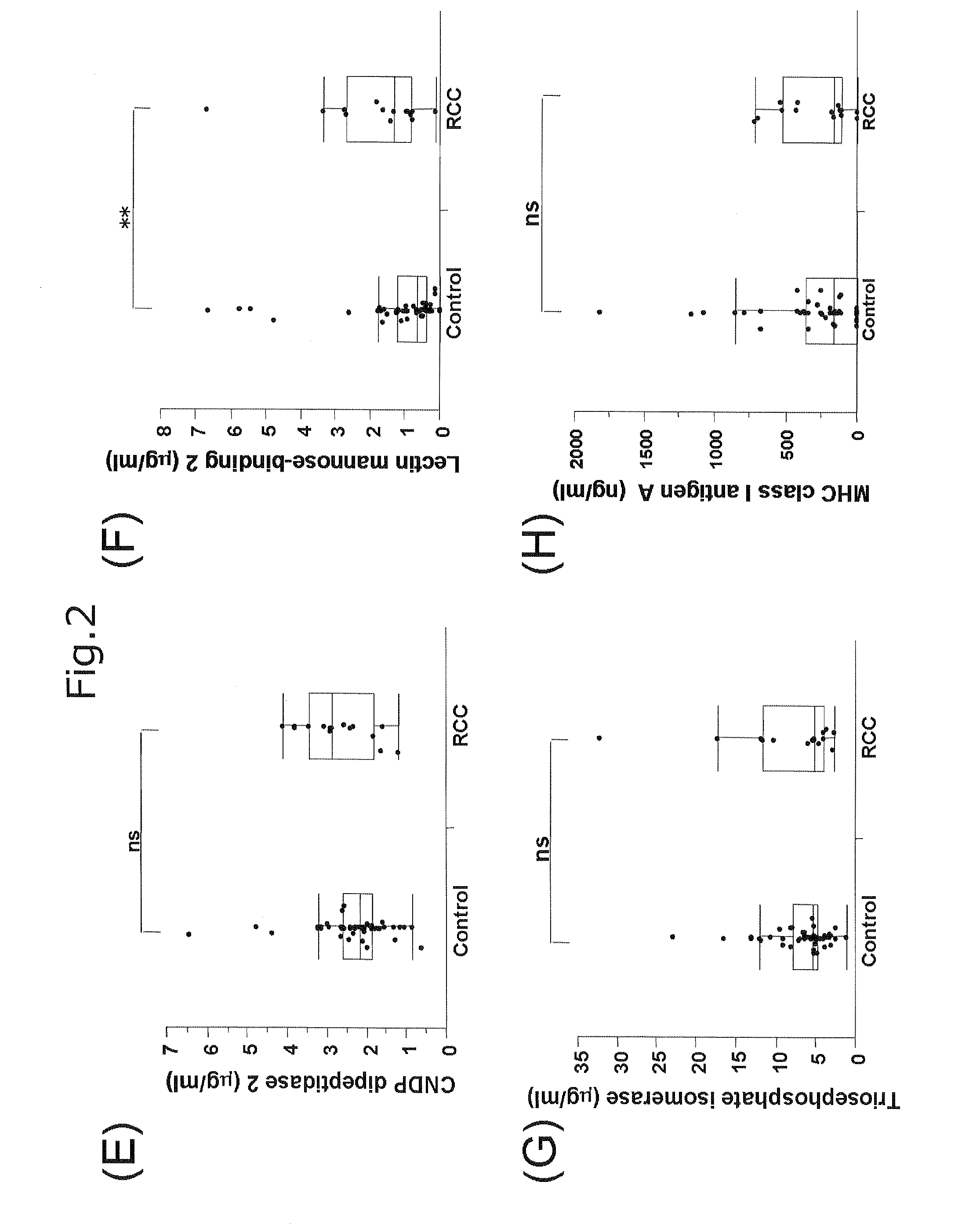
[0069]FIGS. 1 and 2 show graphs each representing the comparison of blood concentration of Glaectin-1 (FIG. 1A), Glaectin-3 (FIG. 1B), α-enolase (FIG. 1C), Ca...
example 3
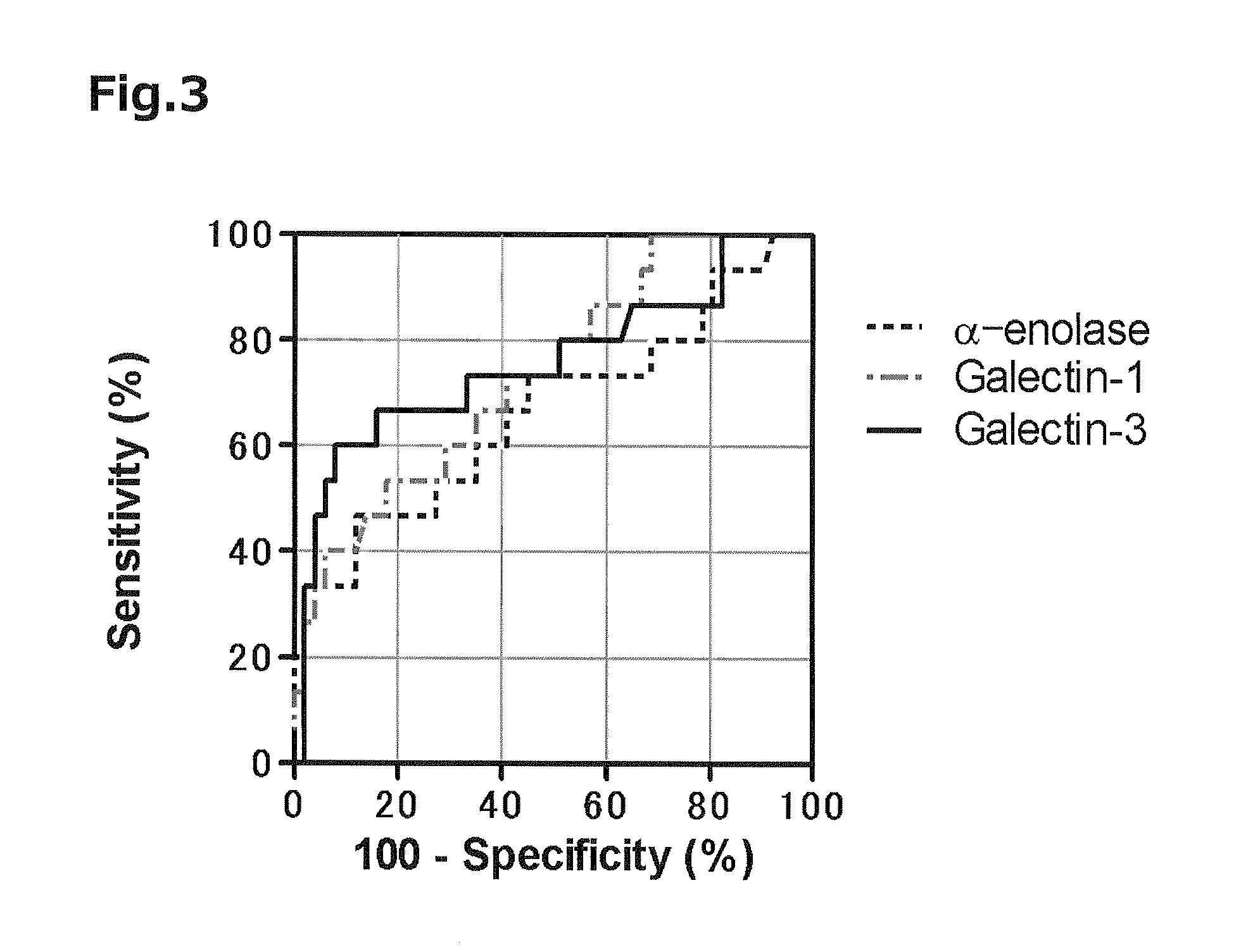
[0076]In order to use the proteins as markers with high accuracy, it is necessary to set their threshold values that indicate both high sensitivity and high specificity. Accordingly, the concentration of each of the proteins that maximized Youden's Index was determined using the ROC curve obtained in Example 2 to set each of threshold values. As a result, a Galectin-1 concentration of 48.4 ng / mL, a Galectin-3 concentration of 18.4 ng / mL, and an α-enolase concentration of 122 ng / mL were derived. At the respective concentration values, Galectin-1 showed a sensitivity of 53.3% and a specificity of 82.4%; Galectin-3 showed a sensitivity of 60.0% and a specificity of 92.2%; and α-enolase showed a sensitivity of 46.7% and a specificity of 88.2%.
[0077]FIGS. 4A to 4C are scatter diagrams each obtained by plotting the concentrations of any two of the three proteins of the renal cancer patients (RCC) and the healthy individuals (Control). In these diagrams, the threshold values set in such a ...
PUM
| Property | Measurement | Unit |
|---|---|---|
| concentration | aaaaa | aaaaa |
| concentration | aaaaa | aaaaa |
| concentration | aaaaa | aaaaa |
Abstract
Description
Claims
Application Information
 - R&D
- Intellectual Property
- Life Sciences
- Materials
- Tech Scout
- Unparalleled Data Quality
- Higher Quality Content
- 60% Fewer Hallucinations
Browse by: Latest US Patents, China's latest patents, Technical Efficacy Thesaurus, Application Domain, Technology Topic, Popular Technical Reports.
© 2025 PatSnap. All rights reserved.Legal|Privacy policy|Modern Slavery Act Transparency Statement|Sitemap|About US| Contact US: help@patsnap.com


