Gradient Porous Scaffolds
a porous scaffold and grade technology, applied in the field of grade porous scaffolds, can solve the problems that the success of fabricating scaffolds for osteochondral repair has been limited, and achieve the effects of increasing porosity, increasing mineralization potential, and increasing porosity
- Summary
- Abstract
- Description
- Claims
- Application Information
AI Technical Summary
Benefits of technology
Problems solved by technology
Method used
Image

Examples
example 1
REFERENCES FOR EXAMPLE 1
[0114]1. Laurencin C, Khan Y, El-Amin S F. Bone graft substitutes. Expert Rev Med Devices. 2006;3(1):49-57.[0115]2. Pneumaticos S G, Triantafyllopoulos G K, Basdra E K, Papavassiliou A G. Segmental bone defects: from cellular and molecular pathways to the development of novel biological treatments. J Cell Mol Med. 2010;14(11):2561-9.[0116]3. Luo J, Sun M H, Kang Q, Peng Y, Jiang W, Luu H H, et al. Gene therapy for bone regeneration. Curr Gene Ther. 2005;5(2):167-79.[0117]4. Laurencin C T, Ambrosio A M, Borden M D, Cooper J A. Tissue engineering: orthopedic applications. Annu Rev Biomed Eng. 1999;1:19-46.[0118]5. Greenwald A S, Boden S D, Goldberg V M, Khan Y, Laurencin C T, Rosier R N, et al. Bone-graft substitutes: facts, fictions, and applications. J Bone Joint Surg Am. 2001;83-A Suppl 2 Pt 2:98-103.[0119]6. Gazdag A R, Lane J M, Glaser D, Forster R A. Alternatives to Autogenous Bone Graft: Efficacy and Indications. J Am Acad Orthop Surg. 1995;3(1):1-8.[012...
example 2
REFERENCES FOR EXAMPLE 2
[0208]Amini, A., D. Adams, C. Laurencin & S. P. Nukavarapu. 2011. Development of Biodegradable Scaffolds with Adequate Porosity and Mechanical Strength for the Regeneration of Segmental Bone Defects (submitted to Tissue Engineering).[0209]Andrassy, M., J. Igwe, F. Autschbach, C. Volz, A. Remppis, M. F. Neurath, E. Schleicher, P. M. Humpert, T. Wendt, B. Liliensiek, M. Morcos, S. Schiekofer, K. Thiele, J. Chen, R. Kientsch-Engel, A. M. Schmidt, W. Stremmel, D. M. Stern, H. A. Katus, P. P. Nawroth & A. Bierhaus (2006) Posttranslationally modified proteins as mediators of sustained intestinal inflammation. Am J Pathol, 169, 1223-37.[0210]Aro, H. T. & A. J. Aho (1993) Clinical use of bone allografts. Ann Med, 25, 403-12.[0211]Arrington, E. D., W. J. Smith, H. G. Chambers, A. L. Bucknell & N. A. Davino (1996) Complications of iliac crest bone graft harvesting. Clin Orthop Relat Res, 300-9.[0212]Balasubramanian, N., P. Bai, G. Buchek, G. Korza & S. K. Weller (2010)...
example 3
Gradient Porous Scaffolds
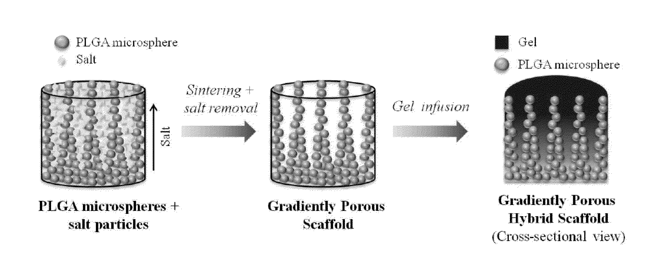
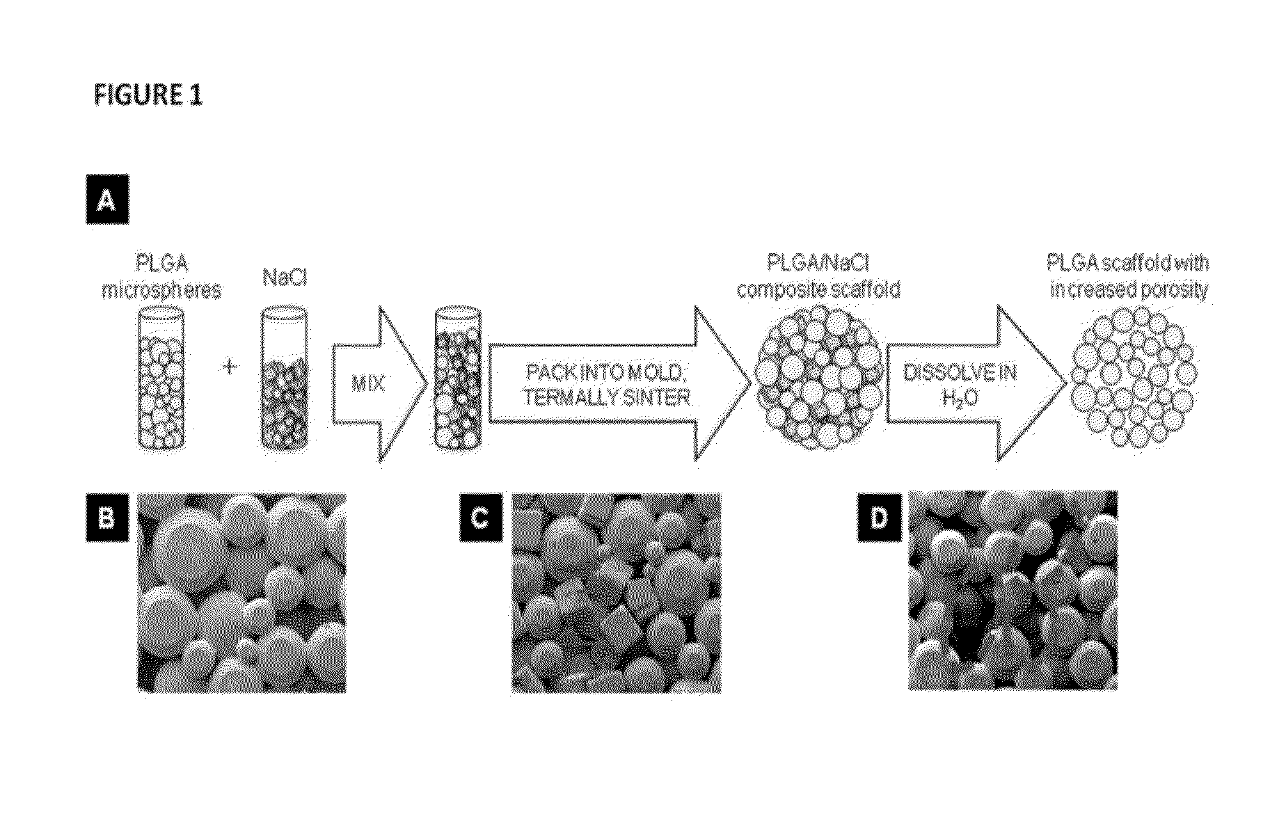
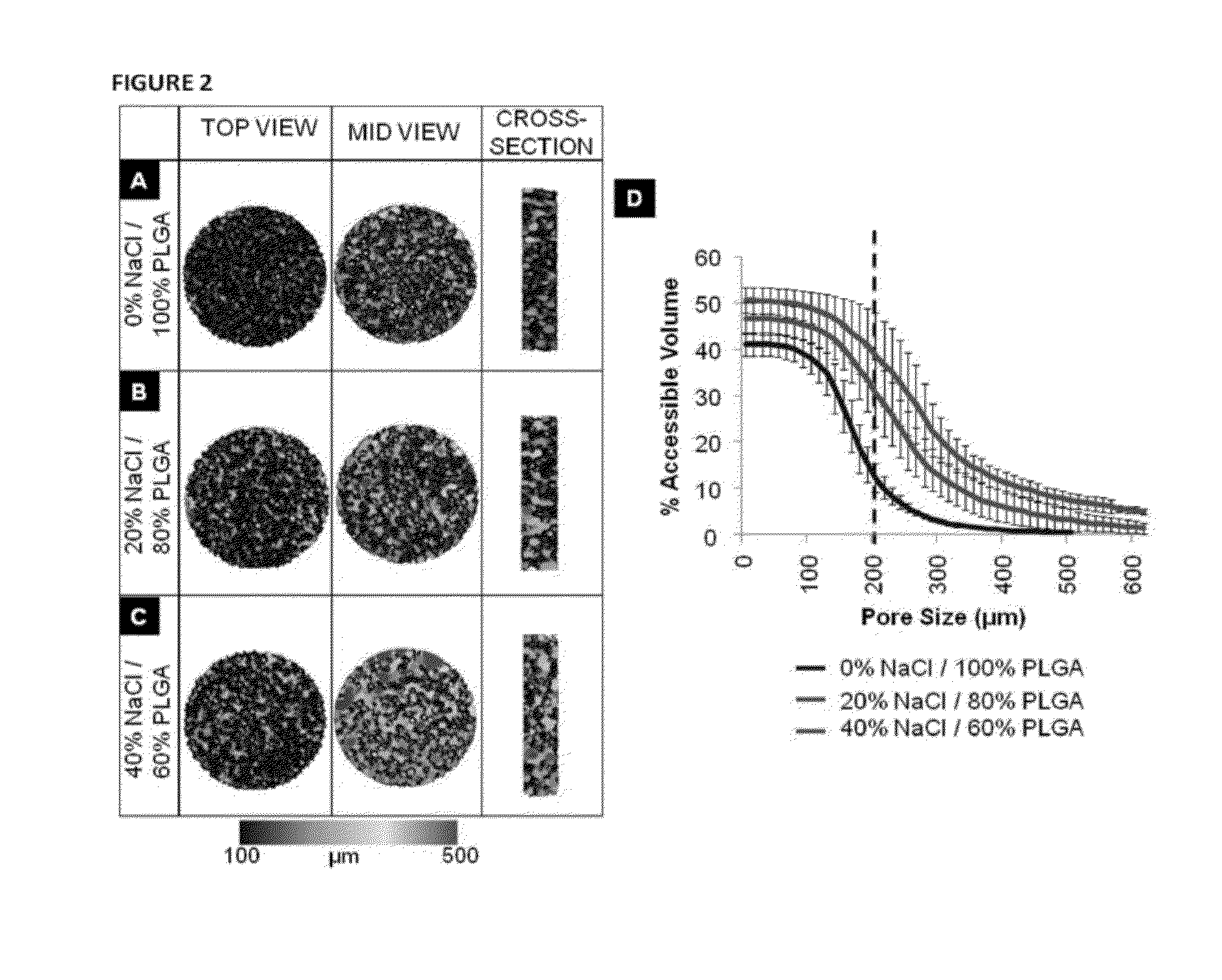
[0251]Articular cartilage is organized into zonal structure (deep, middle, and superficial zones). The deep zone is close to the bony end and mechanically stiff, while the middle and superficial zones consist of soft cartilage. There is gradual change in the amount of collagen and proteoglycans present from deep to superficial zones. To structurally mimic the articular cartilage, we have fabricated novel scaffolds with gradient porosity. The proposed hybrid scaffold with gradient porous structure mimics the native osteochondral structure physically while supporting chondroprogenitor cell survival. The bottom phase of the scaffold extends upwards with larger pore and increasing diameter from bottom to top that mimics the deep to middle zone. An exemplary representation of such a gradient porous scaffold is shown in FIG. 20. Optionally, a self-assembling peptide hydrogel can be used to fill the entire porous structure and constitute the superficial zone (see, ...
PUM
| Property | Measurement | Unit |
|---|---|---|
| Diameter | aaaaa | aaaaa |
| Diameter | aaaaa | aaaaa |
| Diameter | aaaaa | aaaaa |
Abstract
Description
Claims
Application Information
 - R&D
- Intellectual Property
- Life Sciences
- Materials
- Tech Scout
- Unparalleled Data Quality
- Higher Quality Content
- 60% Fewer Hallucinations
Browse by: Latest US Patents, China's latest patents, Technical Efficacy Thesaurus, Application Domain, Technology Topic, Popular Technical Reports.
© 2025 PatSnap. All rights reserved.Legal|Privacy policy|Modern Slavery Act Transparency Statement|Sitemap|About US| Contact US: help@patsnap.com