Topical ocular drug delivery
a technology of ocular drug and composition, applied in the field of topical ocular drug delivery composition, can solve the problems of limited drug permeability across the cornea and conjunctiva, less than 5% bioavailability in anterior segment eye tissue and less than 0.05% in posterior segment eye tissue, and major challenges in intraocular drug delivery, so as to improve the permeability of drug across ocular barriers, increase the effect of drug transporter mediated delivery and poor permeability
- Summary
- Abstract
- Description
- Claims
- Application Information
AI Technical Summary
Benefits of technology
Problems solved by technology
Method used
Image
Examples
example 1
[0057]Determination of Aqueous Solubility of GFX and Ion Pair Complex:
[0058]The aqueous solubility of GFX, GFX-ARG, and GFX-LYS was determined in phosphate buffer saline (PBS) pH 7.4 at 37° C. Solubility was measured by adding an excess amount of GFX (10 mg) to 0.5 ml of PBS containing 1.0 and 3.0 mole equivalent amount of ARG or LYS. PBS without amino acid was included as control. Samples were incubated at 37° C. for 24 hr in incubator shaker with constant shaking at 200 rpm. At the end of 24 hr of incubations, samples were filtered through 0.45 μm filter and filtrates were analyzed for drug content. All experiments were performed in triplicate.
[0059]In Vitro Transport Across Albino Rabbit Cornea and Sclera-Choroid-RPE:
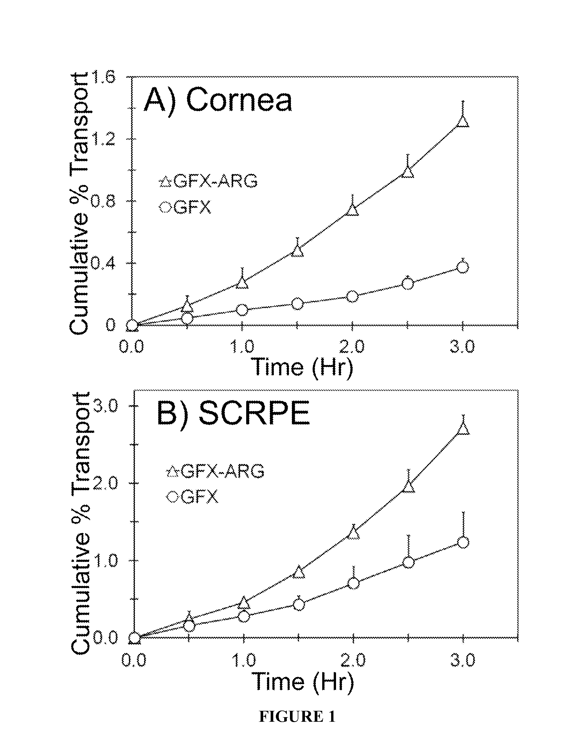
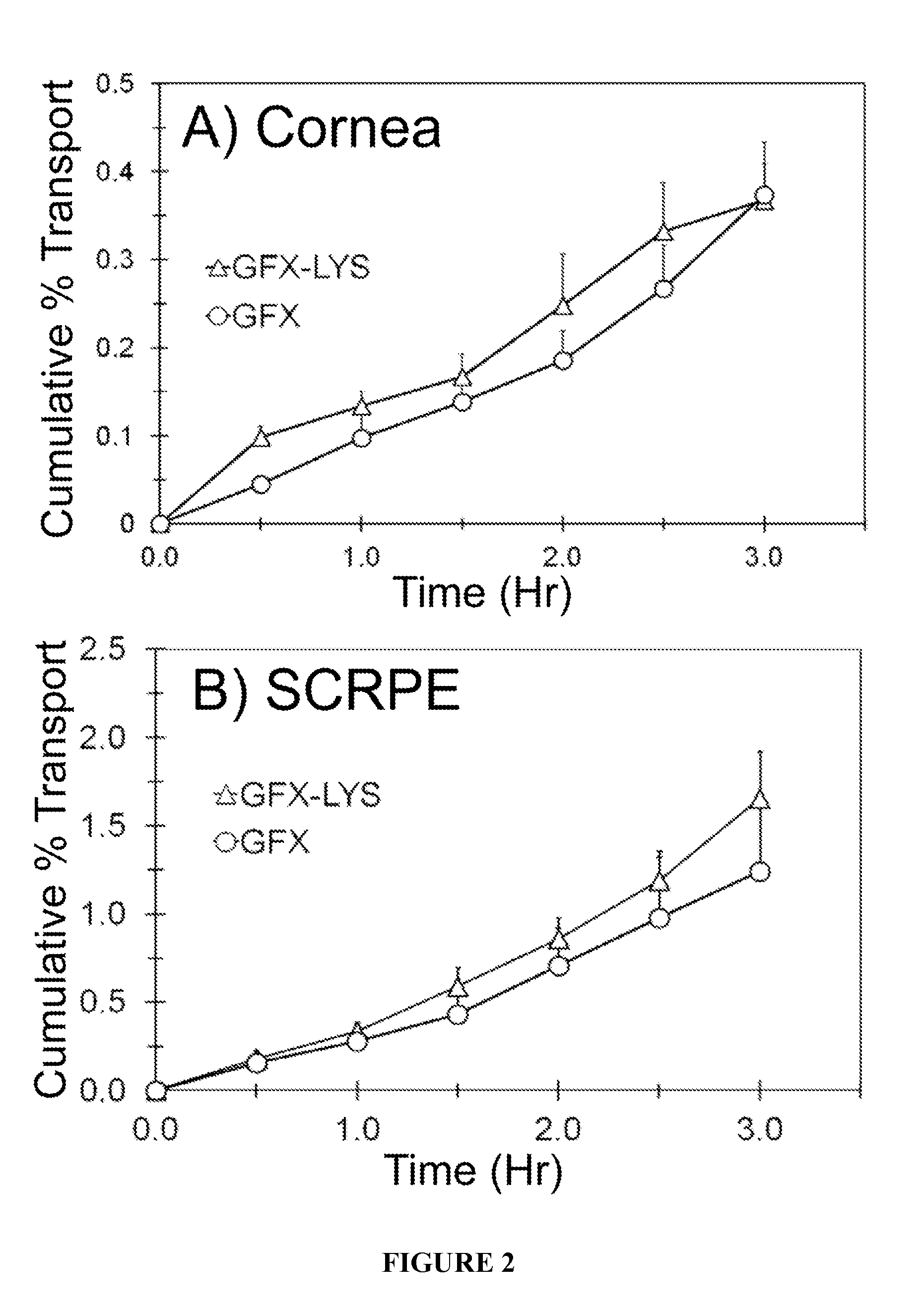
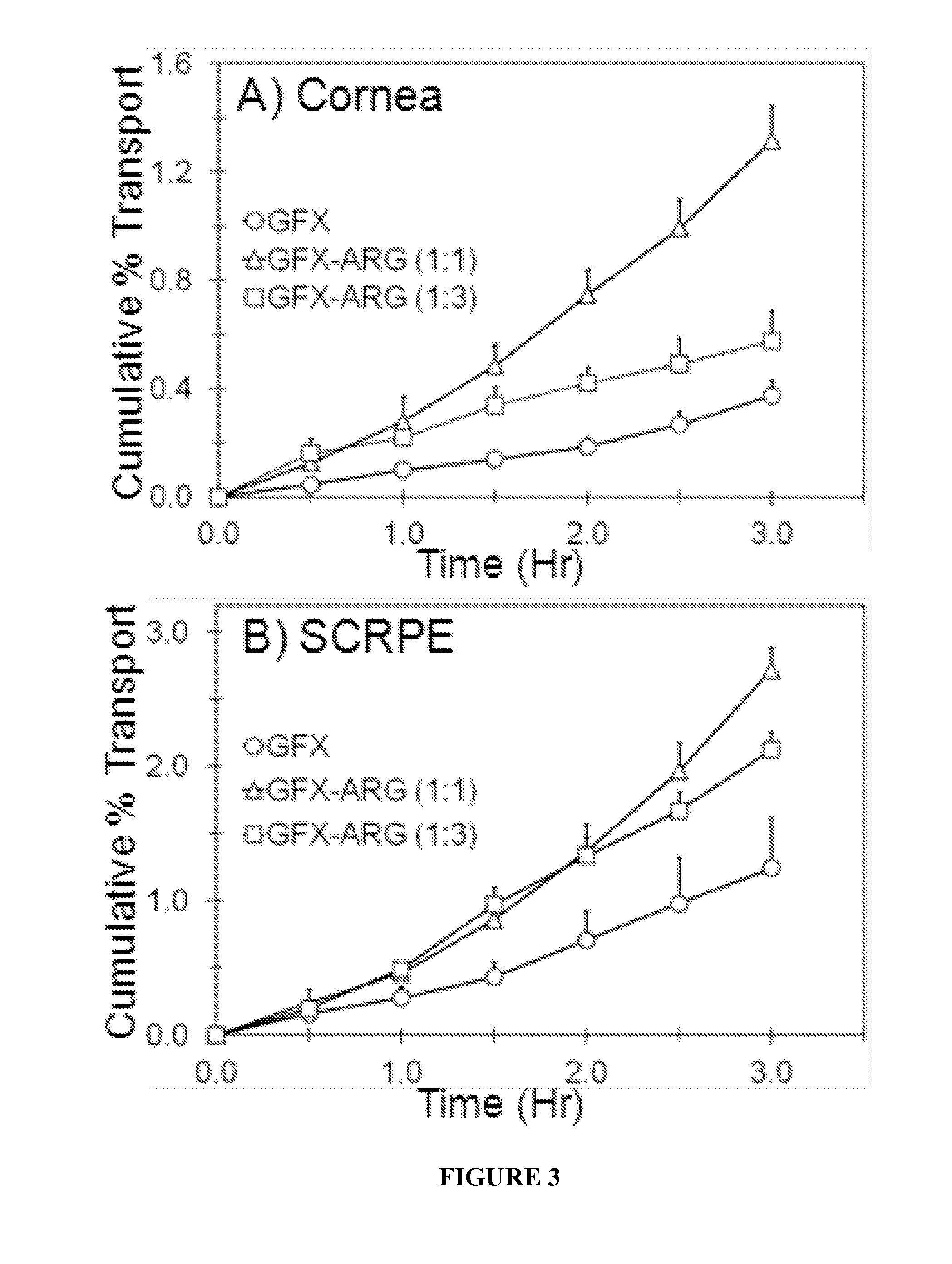
[0060]In vitro transport of GFX, GFX-ARG, and GFX-LYS were carried out across the New Zealand white rabbit cornea and sclera-choroid-RPE (SCRPE). Rabbit eyes were obtained within 24 hr of harvesting from Pel-Freez Biologicals (Rogers, Ark.) and shipped overnight in H...
example 2
[0095]Human Eyes and Tissue Specimens:
[0096]For transport studies, human cadaver eyes were obtained from the Rocky Mountain Lions Eye Bank (Aurora, Colo.) within 48 hrs of death. For immunohistochemical analysis of transporters, human ocular tissue specimens were obtained from archives of University of Colorado, Anschutz Medical Campus eye pathology laboratory. The summary of patient data including age, sex, condition of eye and reason for death are provided in the following Table:
TABLEPatient demographic information.Pa-tientExperimentLensDeathIDperformedSexAgeRaceStatusCause01TransportMale69CaucasianPhakicRenalDisease02TransportFemale56CaucasianPhakicCerebro-vascularAccident03TransportMale65CaucasianPhakicMyocardialInfraction04TransportMale83CaucasianAphakicRenalFailure06IHCFemale52CaucasianPhakicNot known07IHCMale62CaucasianPhakicDiabetes08IHCMale64CaucasianAphakicHeartAttack
For transport study, the eyes were immediately used upon arrival. For immunohistochemistry, formalin-fixed ...
example 3
[0206]While age related macular degeneration (AMD) and diabetic retinopathy are leading causes of blindness in adults, retinopathy of prematurity (ROP) is a leading causes of blindness in infants. Neovascularization of retina and / or choroid is the hallmark of these diseases, with tissue hypoxia being a key cause. Expression of several angiogenic and anti-angiogenic factors are oxygen dependent and controlled by hypoxia inducible factor. Hypoxia stimulates the release of hypoxia induced cytokines including vascular endothelial growth factor (VEGF) that is responsible for retinal neovascularization. Capillary loss in retina or impairment of choroidal blood vessels can result in hypoxia development. Development of hypoxia in choroid / retina stimulates VEGF release, thereby causing choroidal / retinal angiogenesis. In animal models of retinal neovascularization, hypoxia induced VEGF levels correlate with neovascularization.
[0207]Retina is a metabolically active tissue and needs large amoun...
PUM
| Property | Measurement | Unit |
|---|---|---|
| solubility | aaaaa | aaaaa |
| solubility | aaaaa | aaaaa |
| solubility | aaaaa | aaaaa |
Abstract
Description
Claims
Application Information
 - R&D
- Intellectual Property
- Life Sciences
- Materials
- Tech Scout
- Unparalleled Data Quality
- Higher Quality Content
- 60% Fewer Hallucinations
Browse by: Latest US Patents, China's latest patents, Technical Efficacy Thesaurus, Application Domain, Technology Topic, Popular Technical Reports.
© 2025 PatSnap. All rights reserved.Legal|Privacy policy|Modern Slavery Act Transparency Statement|Sitemap|About US| Contact US: help@patsnap.com


