
Integrated assay that combines flow-cytometry and multiplexed HPV genotype identification

a technology of flow cytometry and hpv, which is applied in the field of integrated assays that combine flow cytometry and multiplexed hpv genotype identification, can solve the problems of low specificity, plagued cytological diagnosis of cervical lesions, and inability to identify the underlying high-grade lesions of patients with low-grade cytological abnormalities, so as to achieve enhanced sensitivity and specificity of hpv detection and identification.

Inactive Publication Date: 2013-06-27
PURDUE RES FOUND INC
View PDF3 Cites 2 Cited by
  • Summary
  • Abstract
  • Description
  • Claims
  • Application Information

AI Technical Summary

Benefits of technology

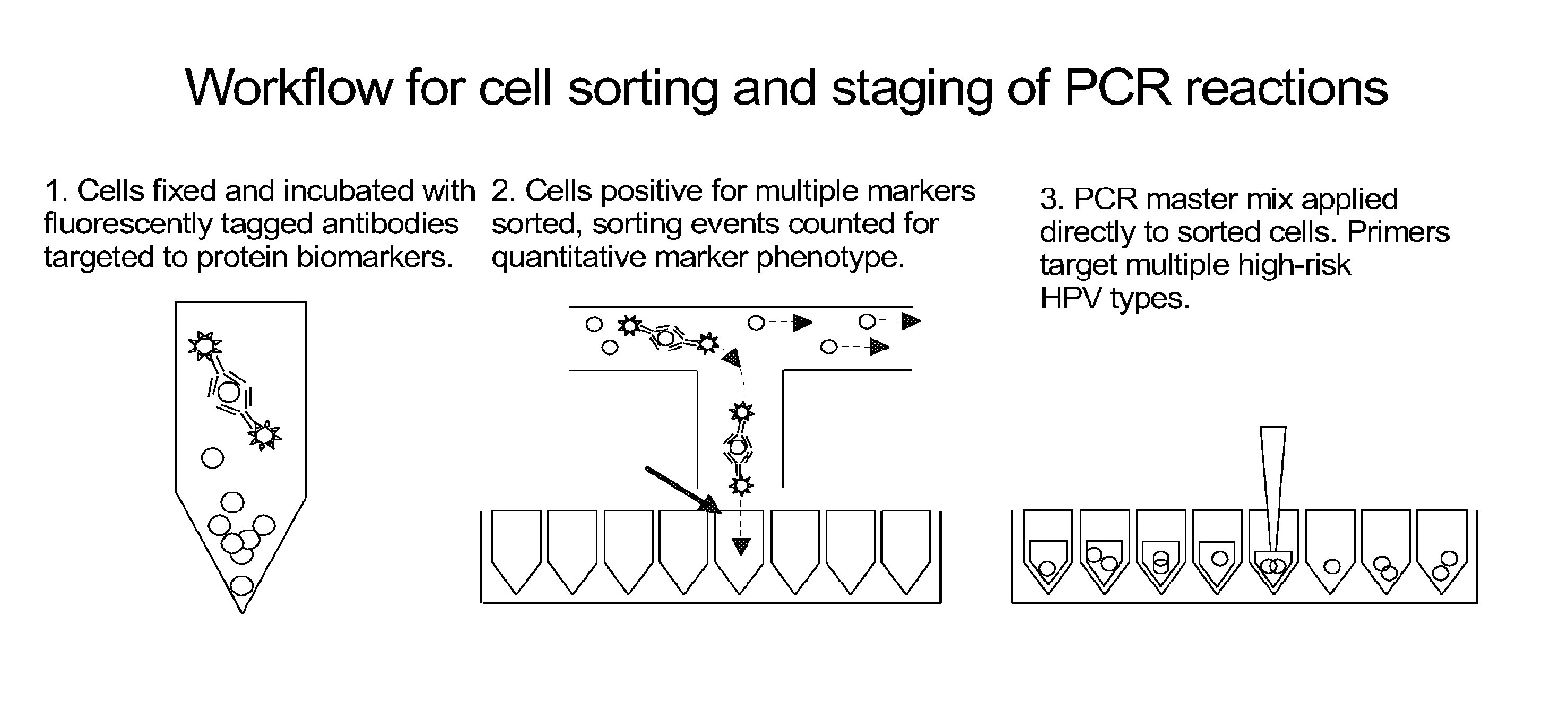
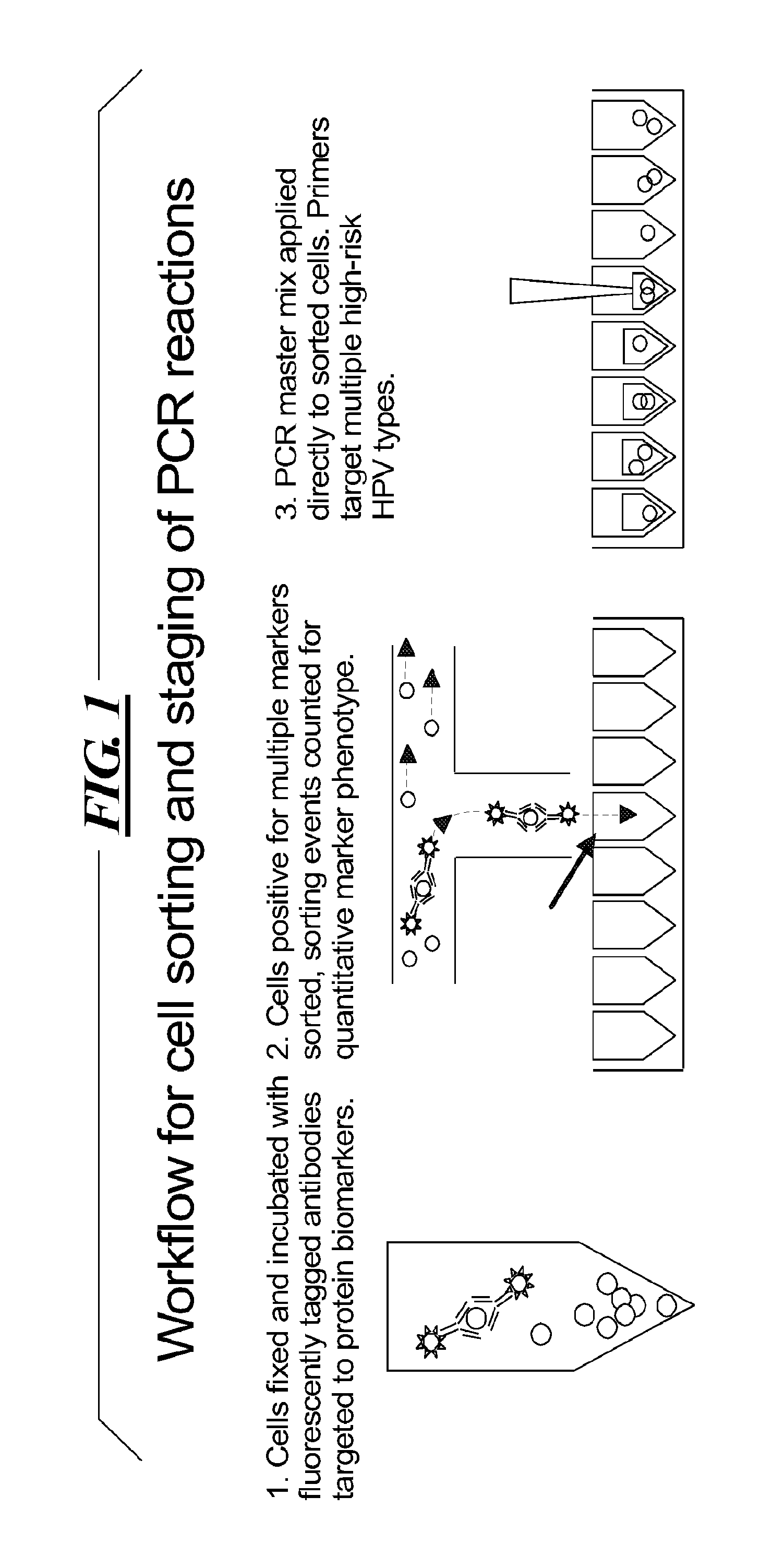
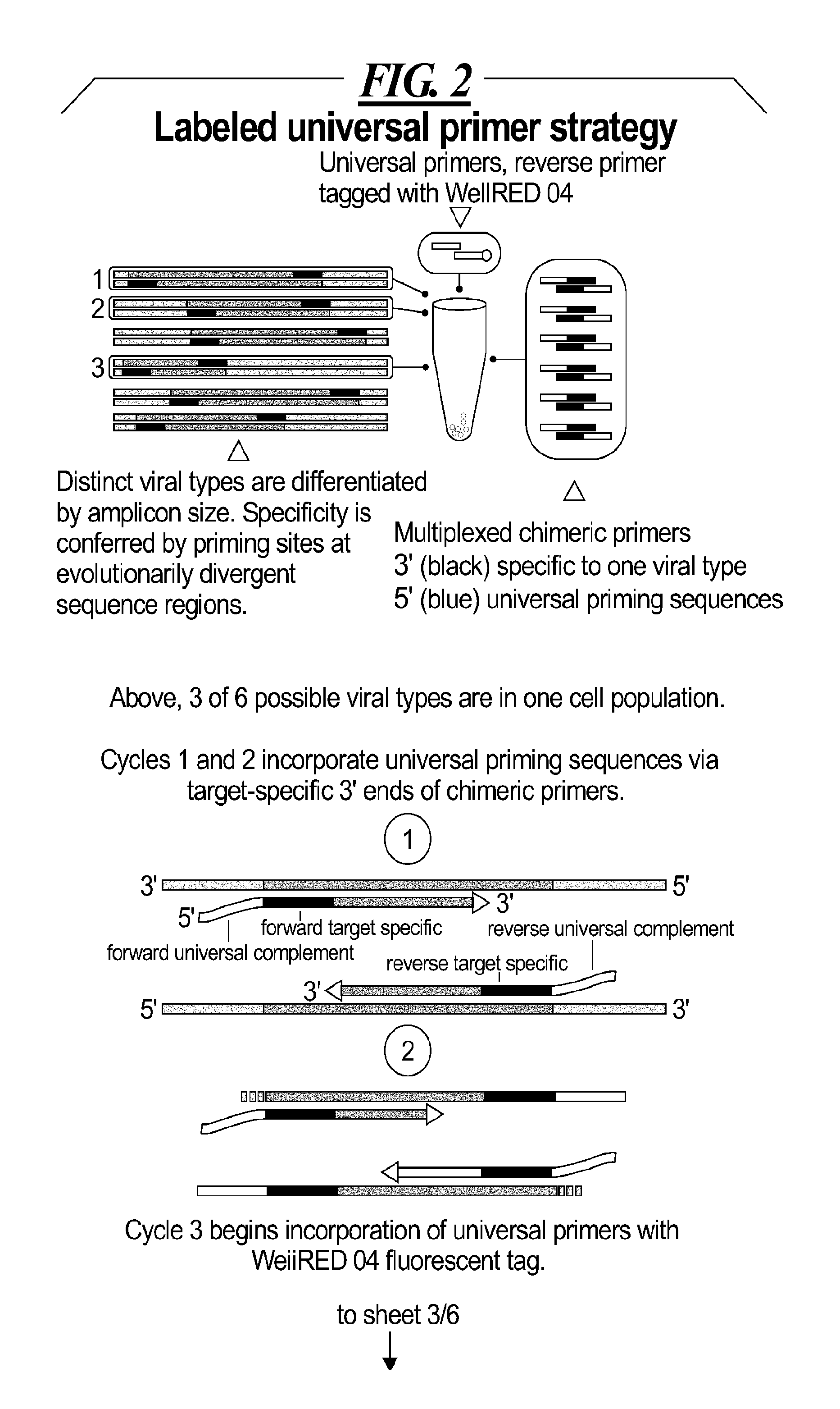
This patent is about a new method that combines two techniques to improve the detection and characterization of cervical disease. The method involves flow-cytometry and identifying specific genotypes of HPV using a two-part assay. The method uses a high-speed cell sorting system to identify cells that have over-expression of specific proteins, which are associated with transformation. These cells are then checked for high-risk HPV genotypes using a set of primers designed to work in a multiplexed reaction. The detection of individual HPV types can be done by measuring the size of amplified fragments using a capillary electrophoresis platform. The method has been optimized to enhance the sensitivity and specificity of detecting and identifying HPV using a thermocycling program that performs denaturation and annealing / extension during very short incubation times for a number of cycles.

Problems solved by technology

Despite the success of Pap tests, the cytological diagnosis of cervical lesions is plagued by a persistent problem of low specificity for clinically significant high-grade lesions in patients with low-grade cytological abnormalities.
In most cases, further evaluation does not identify underlying high-grade lesions in patients with low-grade cytological abnormalities.
Although a simple detection of high-risk types of human papillomavirus can play an important role for the triage of patients, the detection, by itself, has not been useful for several cytological diagnoses.
Specifically, simple detection of high risk HPVs does not predict an underlying high grade lesion because of the long lag time (up to 10 years) between initial infection and development of disease.
Further, some studies indicate that prognosis may vary even among separate high-risk HPV types, yet the current commercially available test for high-risk HPV does not specify individual HPV types.
However, despite its success, the practice of diagnostic cytopathology is limited by poor specificity for underlying clinically significant lesions in cases with low-grade cytological abnormalities.
In most cases, however, further evaluation does not identify high-grade squamous or glandular lesions in patients with low-grade cytological abnormalities.
The above statistics regarding performance of the Pap test of course do not even address the problem of lack of implementation in developing nations, where mortality rate per 100,000 women is 3-7 fold that of more industrialized areas of the world.
Another issue with current practice is that clinicians are under pressure to read large numbers of specimens quickly.
Fatigue and monotony can reduce the acuity of the screener so that rare positive cells have a greater chance of being overlooked.
In pre-cancerous cervical lesions and cervical carcinomas, human papillomavirus DNA integration into the host genome may result in disruption of the viral E2 open reading frame, resulting in unregulated overexpression of HPV oncogenes E6 and E7.
The problem up to this point is that an approximately 10 year latency between infection with high-risk HPV and onset of cancer prevents HPV type data by itself from being prognostically interpreted.
Furthermore, the HC2 test is limited by the absence of an internal control for specimen adequacy.

Method used


Image

  • Integrated assay that combines flow-cytometry and multiplexed HPV genotype identification
  • Integrated assay that combines flow-cytometry and multiplexed HPV genotype identification
  • Integrated assay that combines flow-cytometry and multiplexed HPV genotype identification

Examples


Embodiment Construction

[0035]A two part assay is disclosed that enables collection of both protein biomarker phenotype and specific HPV genotype data from within a clinically derived population of cervical epithelial cells. Data is collected hierarchically. Presence of multiple transformation-associated protein biomarkers acts as a gating criterion for cell sorting, followed by application of a PCR protocol sensitive enough to detect and identify individual HPV types from within the cells captured during sorting. The workflow has been optimized to work with cells conventionally fixed in PreservCyt (Cytyc), and it can be performed on residual cells remaining in a stored sample after a Pap test has been performed.

[0036]Protein biomarker data is quantified proportionally within a cell population by counting positive versus negative sorting events. Thus biomarker data for a clinical sample is not a single value representing an overall staining intensity, but is instead a value reflecting the proportion of the...


PUM

PropertyMeasurementUnit
emission peakaaaaaaaaaa
electrophoresisaaaaaaaaaa
widthaaaaaaaaaa

Abstract

A two part assay is disclosed that enables collection of both protein biomarker phenotype and specific HPV genotype data from within a clinically derived population of cervical epithelial cells. Presence of multiple transformation-associated protein biomarkers acts as a gating criterion for cell sorting, followed by application of a PCR protocol sensitive enough to detect and identify individual HPV types from within the cells captured during sorting. The workflow has been optimized to work with cells conventionally fixed in PreservCyt (Cytyc), and it can be performed on residual cells remaining in a stored sample after a Pap test has been performed.

Description

US GOVERNMENT RIGHTS[0001]This disclosed assays and methods were invented with Government support under Contract No.: R21 CAl25370, awarded by the National Institute of Health (NIH). The U.S. Government may have certain rights to this application.BACKGROUND[0002]1. Technical Field[0003]A two part assay is disclosed that enables collection of both protein biomarker phenotype and specific HPV genotype data from within a clinically derived population of cervical epithelial cells.[0004]2. Description of the Related Art[0005]Cervical cancer is second to breast cancer as the most common form of malignancy in both incidence and mortality for women worldwide. The population-wide utilization of screening cervical cytology (Pap smear tests or “Pap tests”) has been associated with a dramatic decrease in morbidity and mortality from cervical cancer in the United States and in other industrialized nations. Despite the success of Pap tests, the cytological diagnosis of cervical lesions is plagued...

Claims


Application Information

Patent Timeline
Patent Type & Authority Applications(United States)
IPC IPC(8): C12Q1/70
CPCC12Q1/6804C12Q1/708C12Q2600/16C12Q2565/125C12Q2537/143C12Q2531/113
Inventor BIEBERICH, ANDREW A.RAKOTOMALALA, LOVA N.DAVISSON, VINCENT J.ROBINSON, JOSEPH P.
Owner PURDUE RES FOUND INC
Learn More