Mr imaging using navigators

Active Publication Date: 2013-05-23
KONINKLIJKE PHILIPS ELECTRONICS NV
View PDF0 Cites 9 Cited by
  • Summary
  • Abstract
  • Description
  • Claims
  • Application Information

AI Technical Summary

Benefits of technology

The invention is about improving MR imaging by using a method to detect and correct motion during the acquisition process. The method involves applying a unlabeled sequence before the actual MR imaging signals are acquired. This has the effect of reducing the impact of previous imaging sequences and ensuring accurate motion correction. The unlabeled sequence converts the nuclear magnetization in the examined body back into longitudinal magnetization, which prevents saturation bands from interfering with the subsequent imaging sequence. The method also allows for interleaving the acquisition of MR imaging signals and navigator signals, which can be used to perform high-quality MR imaging and spectroscopy even when the navigator and volume of interest are overlapping. In addition, the invention allows for a faster overall scan time by applying the unlabeled sequence and the subsequent navigator sequence without waiting for relaxation of the nuclear magnetization within the volume of interest.

Problems solved by technology

In a variety of MRI applications, motion of the examined object (the patient) can adversely affect image quality.
Motion of the object to be imaged during that finite acquisition time typically results in motion artifacts in the reconstructed MR image.
In the case of medical MR imaging, motion artifacts can result for example from cardiac cycling, respiratory cycling, and other physiological processes, as well as from patient motion.
In dynamic MR imaging scans, the motion of the examined object during data acquisition leads to different kinds of blurring, misregistration, deformation and ghosting artifacts.
Particularly in abdominal applications, the problem arises that the volume of interest, from which the MR imaging signals are acquired, partially overlaps with the navigator volume.
The resulting saturation bands in the MR navigator signals lead to a wrong detection of the contrast edge indicating the position of the diaphragm.
For this reason, the known navigator methods are difficult to apply for MR imaging of the liver or the kidneys.
It can not be avoided that the navigator volume is (at least partly) superimposed upon the respective volumes of interest, with the negative consequence that the image quality is considerably degraded due to the incorrect detection of the respiratory motion state
However, the problem remains that the nuclear magnetization within the volume, from which the MR imaging signals are acquired, remains saturated when the imaging sequence and the navigator sequence are repeatedly applied in an interleaved fashion.

Method used


Image

  • Mr imaging using navigators
  • Mr imaging using navigators
  • Mr imaging using navigators

Examples


Embodiment Construction

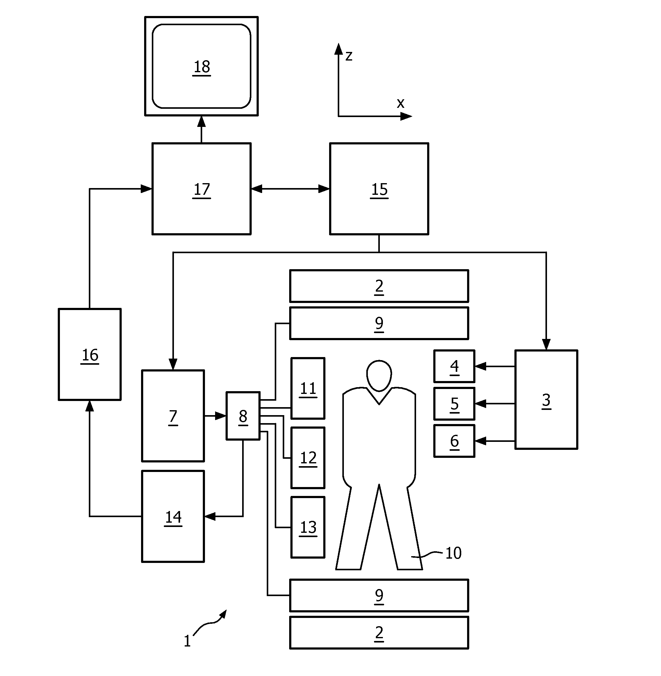
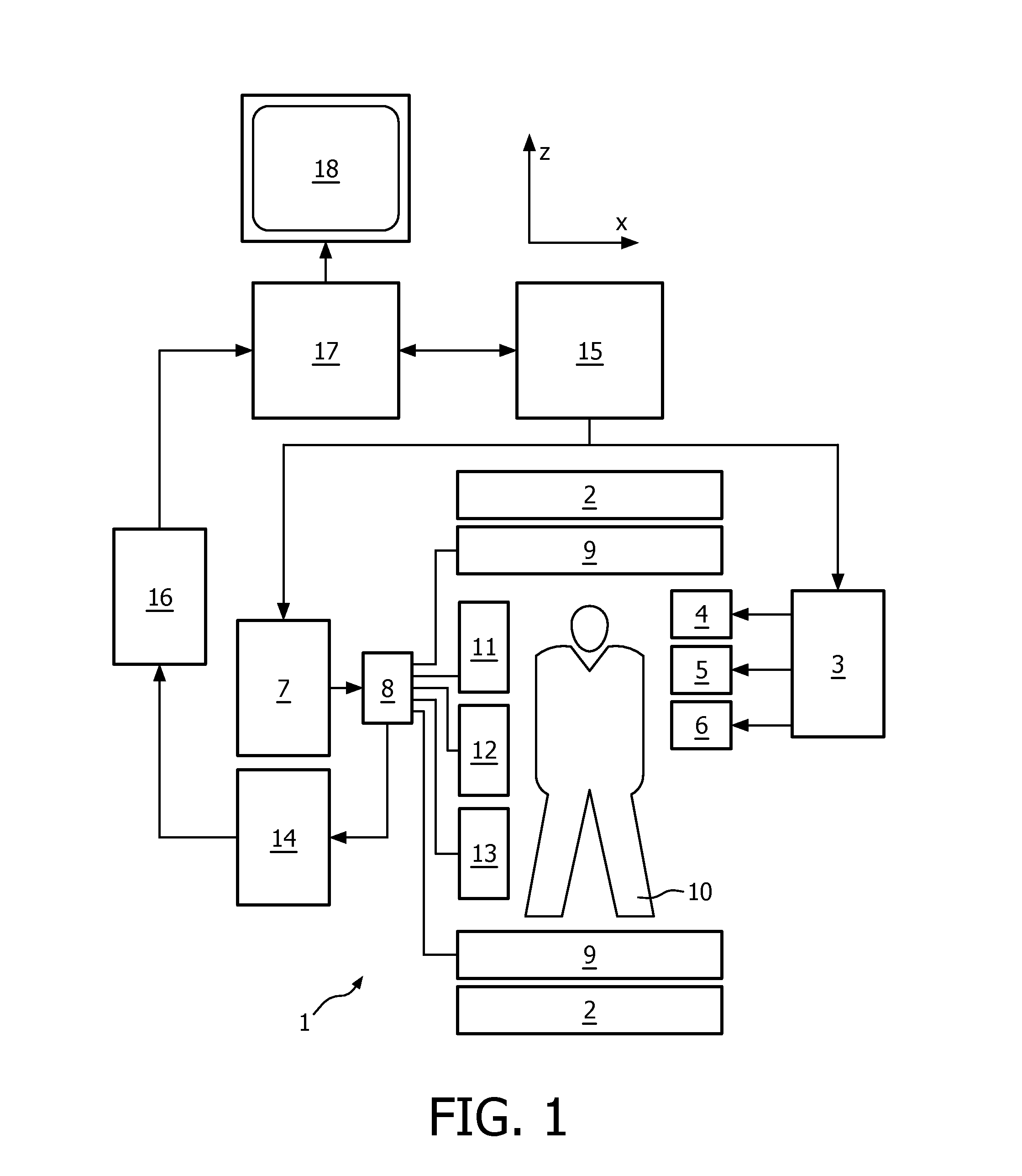
[0038]With reference to FIG. 1, a MR device 1 is shown. The device comprises superconducting or resistive main magnet coils 2 such that a substantially uniform, temporally constant main magnetic field is created along a z-axis through an examination volume.

[0039]A magnetic resonance generation and manipulation system applies a series of RF pulses and switched magnetic field gradients to invert or excite nuclear magnetic spins, induce magnetic resonance, refocus magnetic resonance, manipulate magnetic resonance, spatially and otherwise encode the magnetic resonance, saturate spins, and the like to perform MR imaging.

[0040]Most specifically, a gradient pulse amplifier 3 applies current pulses to selected ones of whole-body gradient coils 4, 5 and 6 along x, y and z-axes of the examination volume. A RF frequency transmitter 7 transmits RF pulses or pulse packets, via a send- / receive switch 8, to a whole-body volume RF coil 9 to transmit RF pulses into the examination volume. A typical ...


PUM


Abstract

The invention relates to a method of magnetic resonance (MR) imaging of at least a portion of a body (10) placed in a stationary and substantially homogeneous main magnetic field. The method comprises the following steps; —exciting nuclear magnetization selectively within a spatially restricted volume of interest (20) by subjecting the portion to an imaging sequence (IMG) comprising at least one RF pulse (α) and switched magnetic field gradients (GX / GY); —acquiring at least one MR imaging signal from the volume of interest (20); —exciting nuclear magnetization within a spatially restricted navigator volume (21) by subjecting said portion to a navigator sequence (NAV) comprising at least one RF pulse and switched magnetic field gradients, wherein the navigator volume (21) at least partially overlaps with the volume of interest (20); —acquiring at least one MR navigator signal from said navigator volume (21); —reconstructing a MR image from the acquired MR imaging signals. It is an object of the invention to enable MR imaging with reliable motion detection and high image quality. For this purpose, the invention proposes that the nuclear magnetization within the volume of interest (20) is transformed back into longitudinal magnetization prior to application of the navigator sequence (NAV) by subjecting said portion to an unlabeling sequence(UNLBL) comprising at least one RF pulse (−α).

Description

FIELD OF THE INVENTION[0001]The present invention relates to the field of magnetic resonance (MR). It finds particular application in conjunction with MR imaging methods and MR devices for diagnostic purposes, and will be described with particular reference thereto. However, it is to be appreciated that the present invention is also amenable to other like applications such as MR spectroscopy.[0002]Image-forming MR methods which utilize the interaction between magnetic fields and nuclear spins in order to form two-dimensional or three-dimensional images are widely used nowadays, notably in the field of medical diagnostics, because for the imaging of soft tissue they are superior to other imaging methods in many respects, do not require ionizing radiation and are usually not invasive.[0003]According to the MR method in general, the body of the patient to be examined is arranged in a strong, uniform magnetic field whose direction at the same time defines an axis (normally the z-axis) o...

Claims


Application Information

Patent Timeline
IPC IPC(8): G01R33/565
CPCG01N24/08G01R33/56572G01R33/5676G01R33/46G01R33/4836G01R33/4838G01R33/543
Inventor BECK, GABRIELE MARIANNE
Owner KONINKLIJKE PHILIPS ELECTRONICS NV
Why Eureka
Try Eureka